Multilingual International Publishing Platform for Humanities and Social Sciences111
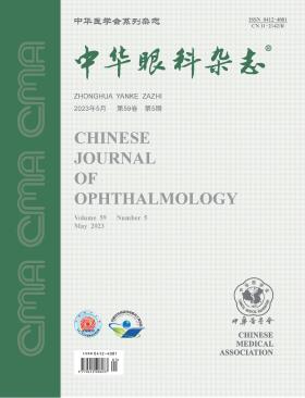
Volume 59 · Issue 05 · 2023
Multilingual International Publishing Platform for Humanities and Social Sciences111
This issue focuses on the research progress of neuro-ophthalmology. The responsible editorial committee responsible for the planning, composition and review of this issue is Professor Zhong Yong.
The optic chiasm is a critical component of the visual pathway, and lesions in the pituitary and sellar regions can cause irreversible damage to a patient′s visual function, resulting in a significant decrease in their quality of life. As a result, neuro-ophthalmology evaluation is a crucial part of the multidisciplinary treatment of pituitary diseases. However, due to the significant variation in the anatomical structure of the optic chiasm and the sellar region, as well as the complexity of the injury mechanism, chiasm injury can result in diverse manifestations and severity levels, which can make it difficult to correlate with anatomical parameters. In recent years, research has increasingly focused on the early recognition of optic chiasm compression, the prediction of visual function after intervention, and the long-term neurodegenerative effects, while optical coherence tomography (OCT), electrophysiological examinations, and functional magnetic resonance imaging are currently the most commonly used methods for evaluating sellar region lesions. However, the role of these methods, represented by OCT, in clinical diagnosis and treatment, still lacks high-level clinical evidence support, and the evaluation and prediction of optic chiasm function remain key areas for further study. In addition to compression lesions, lesions such as inflammation, infiltration, and demyelination in the sellar region, caused by systemic multi-system diseases, can also lead to visual function damage and require recognition in clinical practice.
Papilledema refers to the swelling of the optic disk due to increased intracranial pressure. In clinical practice, papilledema is often confused with optic disc edema. Idiopathic intracranial hypertension (IIH) is a major cause of papilledema and there are still misconceptions about the etiology of IIH, the classification and examination of papilledema, and the treatment of IIH. This article elaborates on the misunderstandings that may exist in the diagnosis and treatment of papilledema, in order to lay a foundation for the standardized diagnosis and treatment of papilledema in China.
Age-related macular degeneration (AMD) is the main cause of low vision and even blindness in the elderly. With the aging of our population, the number of AMD patients will continue to rise. In the past decade, the rapid development of ocular fundus imaging technology has provided a new perspective and approach for the classification, diagnosis and follow-up of AMD. The advent of new drugs has provided more diverse intervention and treatment methods for AMD, especially for neovascular AMD, and the emphasis on accurate and individualized treatment has put forward higher requirements for retinal specialists. Therefore, based on the latest evidence-based medical information, combined with the international guidelines and the current situation of China′s social and economic development, experts from the Chinese Vitreo-Retinal Society of Chinese Medical Association, the Fundus Disease Group of Chinese Ophthalmologist Association, and the National Clinical Research Center for Eye Diseases gave recommendations around eight clinical problems and formed China′s guidelines for the clinical diagnosis and treatment of AMD. With the implementation of these guidelines, we can standardize the diagnosis, treatment, prevention and follow-up of AMD in China. (This article was published ahead of print on the official website of Chinese Journal of Ophthalmology on April 6, 2023)
To investigate the clinical and imaging features of optic nerve tumors that require differential diagnosis from optic neuritis.
A retrospective case series study was conducted. Clinical data of patients diagnosed with optic nerve tumors from January 2017 to December 2021 at the Second Affiliated Hospital of Zhejiang University School of Medicine were collected. A total of 29 patients (39 eyes) with clinical and magnetic resonance imaging (MRI) findings similar to optic neuritis or optic neuropathy were included. There were 15 cases of optic nerve sheath meningioma (ONSM) (17 eyes), 4 cases of optic nerve glioma (ONG) (5 eyes), and 10 cases of infiltrative optic nerve lesions (ION) (17 eyes). All patients underwent best-corrected visual acuity (BCVA), anterior and posterior segment examinations, visual field examination, and orbital or cranial MRI examination. Patient data were observed and analyzed, treatment and follow-up information were recorded, and clinical and imaging features were summarized and compared with those of optic neuritis or optic neuropathy.
Among the 29 patients with optic nerve tumors, 10 were male and 19 were female, with an average age of (43.3±13.8) years and a range of 11 to 72 years. The follow-up time was 6.8 (2.0, 11.0) months, with a range of 1 to 33 months. Sixteen patients (21 eyes) with optic nerve tumors were initially misdiagnosed as having acute optic neuritis and showed poor response to steroid treatment. Of these, 9 cases (11 eyes) were ONSM, 4 cases (6 eyes) were ION, and 3 cases (4 eyes) were ONG. The diagnostic delay time was 7.1 (1.5, 12.0) months, with a range of 1 to 24 months. The main clinical symptoms of all affected eyes were acute vision loss in 23 eyes, slow vision loss in 5 eyes, transient blackouts in 4 eyes, and no obvious visual symptoms in 7 eyes. The median BCVA of all affected eyes was 0.1, ranging from light perception to 1.0. Fundus examination results showed optic disc edema in 34 eyes and normal optic disc in 5 eyes among the 39 eyes with optic nerve tumors. A total of 27 patients (33 eyes) completed visual field examinations, which revealed an enlarged physiological blind spot in 11 eyes, a concentric or tubular visual field contraction in 8 eyes, a diffuse decrease in light sensitivity in 7 eyes, an arcuate scotoma in 4 eyes, and a normal visual field in 3 eyes. All affected eyes completed orbital or cranial MRI examinations, which showed mild optic nerve thickening in 22 eyes, significant thickening in 6 eyes with distortion, and no significant thickening in 6 eyes. Contrast-enhanced T1-weighted imaging (T1WI) MRI showed optic nerve parenchymal thickening in 5 eyes, all of which were ONG, and 2 of them had optic nerve parenchymal enhancement. Optic nerve sheath thickening and enhancement without optic nerve parenchymal thickening or enhancement were observed in 28 eyes, including 17 eyes of ION and 11 eyes of ONSM. There were 6 eyes with no obvious optic nerve thickening, which were all ONSM, showing mild or significant thickening and enhancement of the optic nerve sheath without optic nerve parenchymal thickening or enhancement.
Optic nerve tumors can present with ophthalmic clinical features similar to optic neuritis, such as optic disc edema, and demonstrate MRI findings that resemble those of optic neuritis. Therefore, differentiation between the two is crucial, based on differences in their natural course and response to steroid therapy.
To investigate the characteristics of initial ocular symptoms in children with optic pathway glioma (OPG) at different age stages.
A retrospective case series study was conducted. Clinical data of 16 children with OPG who were diagnosed and treated in the Department of Ophthalmology, Beijing Children′s Hospital, Capital Medical University from April 2017 to July 2021 were collected. The initial ocular symptoms, clinical manifestations of the eyes and nervous system, imaging and histopathological features were analyzed, and the differences in initial ocular symptoms between infants and young children aged≤36 months and older children aged>36 months were compared.
Of all 16 children included, 9 were male and 7 were female. The onset age was 15.0 (6.3, 56.5) months, and the diagnosis age was 48.0 (11.3, 78.0) months. There were 11 infants and young children, and 5 older children. Eye symptoms were the initial manifestation in 11 cases, including 8 cases of infants and young children (5 cases presented with irregular nystagmus, 2 cases with strabismus, and 1 case with failure to chase visual target), and 3 cases of older children (2 cases presented with decreased vision and 1 case with optic disc edema). The imaging findings showed that optic chiasm-involved OPG accounted for the highest proportion, with 3 cases in older children and 8 cases in infants and young children. Among the 8 children with optic chiasm-involved OPG who underwent surgical treatment, 5 were diagnosed with pilocytic astrocytoma according to histopathological results. Seven children had useful vision saved after treatment, and 1 child had visual loss accompanied by cognitive impairment due to surgery after 4 years of nystagmus.
OPG in children often manifests as initial ocular symptoms, with irregular nystagmus being more common in infants and young children, and vision loss being the main symptom in older children.
To investigate the developmental changes of miniature excitatory and inhibitory postsynaptic currents (mEPSCs and mIPSCs) of layer Ⅳ pyramidal neurons in the primary visual cortex binocular zone (V1B) of C57BL/6J wild-type mice at different developmental stages.
Sixteen male C57BL/6J mice of specific-pathogen-free grade were selected and divided into 4 groups according to their postnatal age: P14 group (before and after eye opening), P28 group (the peak of the critical period), P35 group (the end of the critical period), and P130 group (fully adult). Whole-cell patch-clamp technique was used to record the frequency and amplitude of mEPSCs and mIPSCs of layer Ⅳ pyramidal neurons in V1B of each group, and to analyze their differences and changes.
The frequency of mEPSCs of layer Ⅳ pyramidal neurons in V1B of mice in the four groups was statistically different (F=9.46, P<0.001), with the P35 group being higher than the P28 group [P28 and P35 groups were (8.72±1.34) and (13.28±4.05) Hz, t=3.39, P=0.012], and the P130 group being lower than the P35 group [P35 and P130 groups were (13.28±4.05) and (5.82±1.98) Hz, t=5.21, P<0.001]; the amplitude of mEPSCs of layer Ⅳ pyramidal neurons in V1B of mice in the four groups was not statistically different (F=2.84, P=0.055). The frequency of mIPSCs of layer Ⅳ pyramidal neurons in V1B of mice in the four groups was statistically different (F=8.14, P<0.001), with the P130 group being higher than the P14 group [P14 and P130 groups were (5.22±1.33) and (12.03±3.94) Hz, t=4.678, P<0.001]; the amplitude of mIPSCs of layer Ⅳ pyramidal neurons in V1B of mice in the four groups was statistically different (F=7.06, P=0.001), with the P35 group being higher than the P28 group [P28 and P35 groups were (20.07±3.56) and (28.47±5.98) pA, t=3.66, P=0.006], and the P130 group being lower than the P35 group [P35 and P130 groups were (28.47±5.98) and (20.32±3.55) pA, t=3.33, P=0.014].
The excitatory synaptic development of layer Ⅳ pyramidal neurons in V1B of mice is in a vigorous growth state during development and gradually weakens with age, while the inhibitory synaptic development gradually strengthens with increasing postnatal age, and both of them rapidly develop during the critical period.
To investigate the main indications and prognosis of patients undergoing orbital exenteration (OE).
It was a retrospective case series study. The medical records were collected of patients who underwent OE surgery at Beijing Tongren Hospital from January 2007 to December 2021, and their general information, tumor characteristics, lymph node metastasis, diagnosis and treatment, specific surgical techniques, pathological diagnosis and prognosis were analyzed. Kaplan-Meier survival curves were used to evaluate overall survival and recurrence-free survival.
A total of 147 patients were included, with a median age of 58 (48, 68) years. Among them, 82 patients (55.8%) were male and 65 (44.2%) were female, and all underwent unilateral OE. Five patients (3.4%) had benign lesions. Among the 142 patients with malignant tumors, conjunctival malignancies (37.3%) were the most common, followed by eyelid (29.6%), orbital (19.0%), and ocular (14.1%) malignancies. Among the 53 patients with conjunctival tumors, 38 (71.7%) had conjunctival melanoma and 14 (26.4%) had squamous cell carcinoma. Among the 42 patients with eyelid malignancies, 19 (45.2%) had sebaceous gland carcinoma and 16 (38.1%) had basal cell carcinoma. Among the 20 patients with ocular malignancies, 10 (50.0%) had choroidal melanoma and 9 (45.0%) had retinoblastoma. The most common pathological types among the 142 patients with malignant tumors were melanoma (51 cases, 35.9%), squamous cell carcinoma (20 cases, 14.1%), sebaceous gland carcinoma (19 cases, 13.4%), and basal cell carcinoma (16 cases, 11.3%). Of the 135 patients included in the survival analysis, all 5 patients with benign lesions were alive at the last follow-up. The median follow-up time for the 130 patients with malignant tumors was 6.9 (2.5, 6.9) years, ranging from 0.2 to 14.0 years. The overall survival rates at 1, 3, and 5 years after surgery were 90.9% (95%CI: 85.8%-96.0%), 68.4% (95%CI: 59.6%-77.2%), and 60.1% (95%CI: 50.5%-69.7%), respectively. The recurrence-free survival rates of patients with and without lymph node metastasis before surgery were 57.6% and 56.7%, respectively (OR=1.062, 95% CI: 0.525-2.148, P=0.864), and the overall survival rates were 61.5% and 57.7%, respectively (OR=1.019, 95% CI: 0.512-2.033, P=0.957), with no significant differences both. The recurrence-free survival rates of patients with melanoma (47 cases) and non-melanoma (83 cases) were 40.4% and 67.5%, respectively (OR=2.576, 95% CI: 1.390-4.775, P<0.001), and the overall survival rates were 42.6% and 67.5%, respectively (OR=2.845, 95% CI: 1.549-5.225, P<0.001), with significant differences both.
The primary indications for OE are malignant tumors of the conjunctiva and eyelids, with melanoma being the most common malignant tumor of the conjunctiva. Melanoma patients who undergo OE have a lower survival rate compared to other types of malignant tumors. Nevertheless, even in the presence of tumor lymph node metastasis, patients can achieve a relatively good prognosis through OE.
To evaluate the effectiveness and safety of intravitreal dexamethasone implant (IDI) in diabetic macular edema (DME) patients with and without prior vitrectomy.
A retrospective cohort study was conducted on DME patients who received IDI treatment at the Aier Eye Hospital, Beijing from March 2018 to August 2020. Patients were divided into two groups according to whether they had undergone vitrectomy or not. Clinical and follow-up data, including best-corrected visual acuity (BCVA), intraocular pressure (IOP), central macular thickness (CMT), occurrence of ocular and systemic complications, and time to DME recurrence and retreatment, were collected before and after IDI injection at 15 days, 1, 2, 3, and 6 months. Statistical analyses were performed using t-test, Mann-Whitney U-test, χ2 test or Fisher′s exact test, and generalized estimating equations.
Thirty-six patients (41 eyes) were included, with 19 patients (21 eyes) in the vitrectomy group and 17 patients (20 eyes) in the non-vitrectomy group. Compared with baseline, BCVA of eyes in the vitrectomy group was significantly improved at 15 days after IDI injection, with values of 1.00 (0.52, 1.31) and 0.61 (0.30, 1.00), respectively (Z=-2.10, P=0.036); BCVA of eyes in the non-vitrectomy group was significantly improved at 1 month after IDI injection, with values of 0.76 (0.60, 1.35) and 0.52 (0.10, 0.70), respectively (Z=-2.24, P=0.025). Compared with baseline, CMT of eyes in both groups was significantly reduced at all follow-up time points after 15 days of IDI injection (all P<0.05). In the vitrectomy group, CMT before and 15 days after injection were 487 (438, 661) μm and 389 (340, 553) μm, respectively (Z=-3.45, P<0.001); in the non-vitrectomy group, CMT before and 15 days after injection were 486 (410, 641) μm and 323 (290, 396) μm, respectively (Z=-4.07, P<0.001). There were no statistically significant differences in BCVA and CMT between the two groups at all follow-up time points (all P>0.05). The time to DME recurrence was 3.0 (3.0, 4.0) months in the vitrectomy group and 5.0 (4.0, 5.0) months in the non-vitrectomy group, with no significant difference between the two groups (P=0.675). Four eyes (19.0%) in the vitrectomy group and three eyes (15.0%) in the non-vitrectomy group had high IOP, with no significant difference (P=0.529). No severe ocular or systemic complications were observed in any patients.
IDI treatment is safe and effective in DME patients with and without prior vitrectomy, with similar efficacy, but with faster onset of action in patients with prior vitrectomy. There was no significant difference in DME recurrence within 6 months after IDI injection between the two groups.
A 62-year-old female patient presented with binocular vision loss for 10 days. Fundus photography and optical coherence tomography showed bilateral optic disc edema. Fundus fluorescein angiography showed hypofluorescence of the optic disc in the early stage, but irregular filling defects and segmental hyperfluorescence in the late stage. The diagnosis of bilateral simultaneous non-arteritic anterior ischemic optic neuropathy was made. The patient′s visual acuity and visual field were improved after the use of megadose corticosteroids and comprehensive treatment. The prognosis of the patient was stable during the follow-up period.
A 27-year-old male patient had progressive vision loss in both eyes, which was mainly manifested by impaired ganglion cells in the macular area, accompanied by systemic muscle atrophy in limbs. A complete mitochondrial exon gene detection was performed. The final diagnosis was bilateral optic atrophy and axonal Charcot-Marie-Tooth disease 2A2A caused by mutations of the MFN2 gene. There has been no effective treatment. Applications of nutrients to restore the mitochondrial function may alleviate the clinical symptoms.
A 75-year-old male patient presented to the hospital with metamorphopsia of the left eye for 1 month. The corrected visual acuity of left eye was 0.5. Fundus examination showed leopard fundus, small patches of pigmentation were present in the macular area, depigmentation around the macula,choroidal macrovessel emerged from the macular area in a horizontal, temporal, serpentine pattern, optical coherence tomography showed an abnormally enlarged hypo-reflective cavity occupying the full thickness of the choroid in the subfovea as well as in the temporal side of the macula. Early fundus indocyanine green angiography showed rapid filling fluorescence tracks distributed from macula to the temporal side. The patient was diagnosed with macular choroidal macrovessel based on medical history, ocular multimodal examination and general examination. The patient was not given special treatment, and was instructed to control blood pressure in the department of internal medicine and to follow up regularly in the outpatient clinic of fundus disease.
The patient was a 55-year-old male. He was admitted to the Department of Ophthalmology of Xinhua Hospital affiliated to Shanghai Jiaotong University School of Medicine for 8 years due to decreased visual acuity in his right eye and eye pain. Five years ago, he underwent phacoemulsification and cataract aspiration of the left eye combined with anterior peripheral vitrectomy in this hospital. Eye examination: visual acuity is light sensing in the right eye and 0.2 in the left eye; IOP was 52 mmHg (1 mmHg =0.133 kPa) in the right eye and 16 mmHg in the left eye; Adhesive deformation of the right eye pupil with a small amount of new blood vessels, opacity of the lens, and unclear vision of the fundus; The left eye has a round pupil, absent lens, clear and white optic disc boundary, flat retina, sunset-like fundus changes, scattered Dalen-Fuchs nodules in the peripheral retina, and 6 vortex veins are clearly visible (Quintessential Picture 1). Clinical diagnosis: Fogt-Koyanagi-Harada syndrome (both eyes).
The patient was a 46-year-old female. He was admitted to the Department of Ophthalmology of Peking University International Hospital because his left eye visual acuity decreased for 3 months with orbital pain and aggravated for 2 weeks. Eye examination: naked eye visual acuity was 1.0 in the right eye and 20 cm manual in the left eye; IOP was 10 mmHg (1 mmHg =0.133 kPa) in the right eye and 12 mmHg in the left eye; Transparent cornea in both eyes, no retrocorneal sediment, deep anterior chamber, no anterior chamber flash, clear iris texture, round pupil, right eye pupil diameter 3 mm, sensitive to light, left eye pupil diameter 4 mm, showing relative afferent pupil dysfunction, transparent lens; There was no obvious abnormality in the retina of the right eye, edema above the nose of the optic disc and atrophy below the temporal in the left eye, ciliary shunt of the optic nerve on the surface of the optic disc, linear hemorrhage above the temporal of the optic disc, and no obvious abnormality in the macular area. Fluorescein fundus angiography of the left eye is shown in the essence picture 1. Enhanced orbital MRI examination: the optic nerve of the left eye showed a "double track" sign (Quintessential Picture 2), and space-occupying lesions were seen in the optic nerve sheath and anterior clinoid process. Clinical diagnosis: meningioma of the optic nerve sheath of the left eye.
Optic nerve glioma (ONG) is a relatively rare central nervous system tumor that mainly affects children and adolescents. It can be classified into sporadic and neurofibromatosis type 1 (NF1)-associated types. The histological type is mainly a low-grade pilocytic astrocytoma. The typical clinical manifestations are visual impairment and painless eye protrusion, and the imaging features mainly present as fusiform swelling or irregular masses. Chemotherapy is still the first-line treatment for ONG, and other treatment options include radiotherapy, surgical resection, and molecular targeted therapy. Screening and monitoring of NF1 patients are also crucial. The prognosis of ONG is difficult to predict, and close monitoring and timely effective intervention are necessary.
Anterior chamber paracentesis (ACP) is a commonly used ophthalmic technique with broad clinical applications. It plays an important role in the diagnosis of intraocular infections, vitreoretinal lymphoma, and other diseases. However, the current ACP methods used in clinical practice have some issues and potential risks. There is no unified standard for the procedural steps, environment, and perioperative management. Therefore, this article provides a systematic review of various methods and devices for ACP and briefly summarizes the current clinical ACP environment, perioperative management, and safety considerations. The aim of this review is to provide insights into the current status and future prospects of ACP, with the goal of optimizing the safety and efficacy of this technique.
Non-arteritic anterior ischemic optic neuropathy (NAION) is the most common acute optic neuropathy in people over 50 years old. It is caused by hypoperfusion of the short posterior ciliary artery, which leads to insufficient blood supply to the optic nerve in front of the sieve plate, and irreversible large-scale visual field defect caused by ischemic necrosis of optic nerve fibers. At present, there is no effective treatment and poor prognosis. Therefore, prevention of NAION is an urgent task. This article expounds the local and systemic risk factors and susceptibility genes of NAION, hoping to help ophthalmologists deeply understand the risk factors of NAION, carry out in-depth research on the pathogenesis of NAION, and provide effective prevention and treatment measures for patients.
CURRENT ISSUE