Multilingual International Publishing Platform for Humanities and Social Sciences111
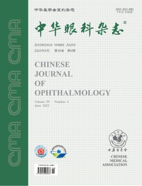
Volume 59 · Issue 06 · 2023
Multilingual International Publishing Platform for Humanities and Social Sciences111
This issue focuses on the research progress of optometry and refractive surgery. The responsible editorial committee responsible for the planning, composition and review of this issue is Professor Wang Yan.
Myopia prevention and control has become one of the difficulties of eye health in China. Previous studies have focused on the effect of myopia control, but less on the visual performance and visual quality caused by various approaches to myopia control for adolescents. On one hand, most of the prevention methods can achieve myopia control by affecting retinal imaging. On the other hand, they can affect the visual quality and even the quality of daily life. Therefore, a balance between myopia control effects and visual quality needs to be considered in clinical work. This article points out the importance of improving visual quality in the process of myopia prevention and control, analyzes the problems of visual quality related to the myopia prevention and control methods, and puts forward suggestions on myopia management, so as to improve the visual quality with the effective prevention and control of myopia in adolescents.Finally, improving the comprehensive prevention and control level of myopia for adolescents.Finally, improving the comprehensive prevention and control level of myopia for adolescents.
The objective of this retrospective study was to employ machine learning techniques to examine age-related traits of ocular aberrations in a substantial population with myopia and myopic astigmatism.
This was a cross-sectional study. Data from a population of myopic and myopic astigmatism patients who underwent wavefront aberration examinations at the Refractive Surgery Center of Tianjin Eye Hospital in Tianjin, China, were collected continuously from January 2013 to July 2017. The data from the right eye of each individual were collected for analysis. Each eye had 32 outcome data points, including age, best-corrected visual acuity (BCVA), lower-order aberrations (spherical diopter, cylindrical diopter, and astigmatic axis), and higher-order aberrations [Zernike coefficients and root mean square (RMS) of the third to sixth order aberrations] were analyzed. Higher-order aberrations were measured by Hartmann-Shack aberrometer.
The study included 1 507 subjects (1 507 eyes), comprising of 694 males and 813 females, with a mean age of (23.28±5.45) years. The findings demonstrated a decrease followed by an increase in most of the higher-order aberrations with age between 15-40 years. The minimum value points were observed in the age group of 25-30 years for RMS of total higher-order aberrations, 3rd RMS (with a confidence of 47.74% for range 1), 4th RMS (with a confidence of 86.01% for range 1), and trefoil aberrations (with a confidence of 56.38% for C33 and 73.25% for C3-3). The minimum value points were also observed in the age group of 30-35 years for primary spherical aberration (with a confidence of 56.10% for C40) and vertical coma-like aberration (with a confidence of 56.91% for C3-1). In contrast, astigmatism with the rule tended to decrease with age, while astigmatism against the rule and oblique astigmatism tended to increase (with a confidence of 88.66%, 84.71%, 81.07%, 79.67%, and 66.35% for astigmatism with the rule in different age groups).
As age increases, the population with with-the-rule astigmatism decreases while the population with against-the-rule astigmatism increases. The high-order aberrations are the lowest in the 25-35 age group.
To explore the characteristics of orthokeratology lenses designed for small correction zones and their effectiveness and safety in long-term prevention and control of myopia in children and adolescents.
It was a prospective cohort study that included myopic children and adolescents who received corneal reshaping treatment at the Qingdao Eye Hospital of Shandong First Medical University between January 2019 and May 2020. The patients were randomly divided into two groups using computer-generated randomization, and were fitted with corneal reshaping lenses with small treatment zones and conventional designs, respectively. The uncorrected visual acuity, best-corrected visual acuity, refractive error, and axial length were measured before and after 6, 12, and 18 months of wearing the lenses. Corneal topography with the Pentacam was also performed, and the area and diameter of the corneal treatment zone were calculated using the Matlab software.
A total of 60 myopic patients (60 eyes) were enrolled, including 29 males and 31 females, with an age of (10.40±1.01) years and a spherical equivalent of (-2.88±0.42) D. There were 30 cases in the small correction zone group and 30 cases in the conventional group. There was no significant difference in uncorrected visual acuity and spherical equivalent between the two groups at each time point after treatment. The axial growth in the conventional group was (0.16±0.09) mm at 6 months after treatment, (0.28±0.17) mm at 12 months, and (0.37±0.20) mm at 18 months, whereas in the small treatment zone group it was (0.06±0.05) mm, (0.12±0.10) mm, and (0.18±0.14) mm, respectively. The myopia progression rate in the small treatment zone group was only 37.50%, 42.86%, and 48.64% of the conventional group at 6, 12, and 18 months, respectively. Corneal topography showed that the treatment area in the conventional group was (6.98±0.89) mm², while in the small treatment zone group it decreased by 23.2% [(5.36±0.73) mm²] (P<0.05). Correlation analysis revealed that the axial increase after 18 months of lens wearing was negatively correlated with the age before lens wearing (P<0.05), positively correlated with the corneal surface treatment zone size (P<0.05), and not correlated with the pupil diameter and spherical equivalent (all P>0.05). After the shaping treatment, the rate of adverse reaction, which was mild, in both groups was 10%, and the symptoms disappeared.
Orthokeratology lenses with smaller treatment zones can significantly reduce the rate of axial length growth in children and adolescents compared to lenses with conventional treatment zones, without compromising treatment safety.
To design a visual fatigue questionnaire that can be used for population surveys.
This was a cross-sectional study that involved three stages of subjects′ recruitment. In the first stage, by convenience sampling, 150 individuals who complained of visual fatigue were selected at public places in Wenzhou City in May 2016. The 19-Item Asthenopia Survey Questionnaire (ASQ-19) was used to conduct the survey, and the questionnaire was adjusted. In the second stage, 200 outpatient participants were recruited from Wenzhou Medical University Affiliated Eye and Optometry Hospital from June 2016 to May 2017 and were divided into a visual fatigue group and a control group based on clinical diagnosis. The adjusted visual fatigue questionnaire was used for validation. In the third stage, 64 outpatient participants who met the inclusion criteria were continuously recruited from the Wenzhou Medical University Affiliated Eye and Optometry Hospital in July 2022. They were tested using the adjusted visual fatigue questionnaire and retested one week later. During the questionnaire adjustment stage, factor analysis and feedback were used to adjust the scoring method and items of the ASQ-19 questionnaire. The adjusted questionnaire was then analyzed for reliability, validity, accuracy, and subject acceptance during the validation and retest stages.
A total of 403 participants were included, and 456 questionnaires were distributed. Eventually, 432 valid questionnaires were collected from 379 participants, resulting in a valid response rate of 94.7%. During the questionnaire adjustment phase, there were 140 valid questionnaires from 140 participants consisting of 56 males and 84 females with an average age of (35.2±12.4) years. In the questionnaire validation phase, there were 186 valid questionnaires from 186 participants. Sixty-two participants had visual fatigue and 124 were controls. During the questionnaire retesting phase, 53 participants yielded 106 valid questionnaires. The group consisted of 20 males and 33 females with an average age of (22.8±4.9) years. After factor analysis, the symptom severity graded as none, mild, moderate, severe, and very severe was scored as 0, 1, 2, 3, and 4 points, respectively. The total score was 44, and the final questionnaire consisted of 11 items (numbered 1, 2, 3, 5, 6, 8, 10, 15, 17, 18, and 19). The 11-Item Asthenopia Survey Questionnaire (ASQ-11) had a Cronbach′s α coefficient of 0.89, a split-half reliability of 0.82, and a test-retest Pearson correlation coefficient of 0.90 (P<0.001). The structural validity was 51.26%, and the discriminative validity was a t-value of 9.19 (P<0.001). On average, it took (2.82±0.43) minutes for participants to complete the questionnaire. The receiver operating characteristic curve had a cutoff value of 8.5, with a sensitivity of 74.19% and a specificity of 80.65%.
The ASQ-11, with fewer items and a shorter completion time, is easy for participants to use and is suitable for screening or self-assessment of visual fatigue in the general population. Additionally, it is convenient for clinical and epidemiological studies related to visual fatigue.
To compare the accuracy of different corneal curvature parameters in assessing the corneal refractive status and tracking corneal power changes after small incision lenticule extraction (SMILE).
This prospective cross-sectional study tracked and recorded total corneal curvature parameters measured by different instruments before and three months after SMILE for myopia. These parameters, including total keratometry (TK) from the IOLMaster 700, total corneal refractive power (TCRP) from the Pentacam AXL, real keratometry (RK) from the CASIA 2, and corrected parameters calculated using the Haigis, Shammas, and Maloney methods, were compared with data obtained using the clinical history method (CHM). Surgically induced changes in TK, TCRP, and RK were analyzed and compared with those in spherical equivalent on the corneal plane (ΔSEco).
The study included 40 eyes (40 participants). After SMILE, the difference was smallest between TK [(0.08±0.38) D] and CHM values (P>0.05). However, TCRP, RK, KHaigis, KShammas, and KMaloney were significantly different from CHM data (P<0.05). The width of the 95% limits of agreement of TK (1.49 D) was narrowest, followed by that of RK (1.57 D). Pearson analysis showed that each parameter had a good correlation with CHM data. The differences between the changes in TK, TCRP and RK caused by surgery and ΔSEco were (0.03±0.39) D, (0.17±0.43) D, and (-0.19±0.46) D, respectively. The width of the 95% limits of agreement of ΔTK (1.54 D) was narrowest, and the correlation coefficient of ΔTK (0.951) was highest.
The parameter TK of the IOLMaster 700 can provide accurate and objective corneal power evaluation after SMILE.
To analyze the clinical features and treatment outcomes of eyes with contact lens-induced limbal stem cell deficiency (CL-iLSCD).
This cross-sectional study involved patients diagnosed with CL-iLSCD at the Eye, Ear, Nose & Throat Hospital of Fudan University between October 2018 and September 2022. A total of 17 patients (25 eyes) with a mean age of (36.4±6.9) years were enrolled. Among them, 14 were females (82.4%). Corneal and limbal abnormalities, especially the range of epitheliopathy, were observed under a slit lamp biomicroscope with fluorescein staining. Anterior segment optical coherence tomography and in vivo laser scanning confocal microscopy were performed to obtain the central corneal epithelial thickness, density of basal epithelial cells and corneal nerve fiber length. The clinical features of CL-iLSCD, along with their treatment outcomes and related risk factors, were analyzed.
All patients wore soft contact lenses, with an average daily wearing time of (10.5±2.5) hours and a median wearing duration of 10 (4 to 30) years. Ocular symptoms, including decreased vision, ocular discomfort or pain, redness, and photophobia, were present in 22 eyes (88.0%). The most characteristic clinical sign of CL-iLSCD was comb-or whorl-pattern late fluorescein staining under cobalt blue light, which was most commonly seen at the superior limbus (25/25, 100.0%). Additionally, reductions in central corneal epithelial thickness, basal cell density, and corneal nerve fiber length were observed. A comprehensive score was assigned to each eye based on clinical findings and in vivo imaging biomarkers. LSCD was mild, moderate, and severe in 5, 11, and 8 eyes, respectively. A history of misdiagnosis was found in 20 eyes (80.0%). After discontinuing the use of contact lenses and receiving medical treatment, significant improvement was observed in all eyes, with 13 eyes fully recovered.
The symptoms and clinical signs of CL-iLSCD can be subtle at the early stage. Discontinuing contact lens wear and medication are effective to treat CL-iLSCD.
A 22-year-old male presented with complaints of blurred vision in his right eye over the past 2 years following small incision lenticule extraction (SMILE) surgery conducted 4 years ago. Following a thorough ocular examination and evaluation, he was diagnosed with corneal ectasia in the right eye after the SMILE procedure. Subsequently, the patient underwent corneal cross-linking (CXL) treatment in the right eye to prevent the progression of the condition. After 3 months of treatment, the corneal ectasia remained stable. This article outlines the process of diagnosis and treatment, reviews the corneal conditions prior to the SMILE surgery, and analyzes the possible reasons behind the occurrence of postoperative corneal ectasia.
A 50-year-old female patient presented to the Beijing Tongren Eye Center, Beijing Tongren Hospital, Capital Medical University, Beijing Institute of Ophthalmology, with complaints of right eye pain, tearing, and difficulty opening the eye for over a month after intrastromal corneal ring segment (ICRS) implantation 18 years prior in both eyes. Slit lamp examination revealed corneal stromal melting around the ICRS at the 3 to 4 o′clock position of the right eye, with fluorescein staining. Optical coherence tomography showed epithelial and superficial stromal layer defects in the area of the lesion. The patient was diagnosed with corneal melting after ICRS implantation in the right eye. Under general anesthesia, the corneal stromal ring was removed, and deep lamellar keratoplasty was performed. The patient had no discomfort and the corneal graft remained transparent after the surgery.
Oliver-McFarlane syndrome is a rare genetic disorder characterized by long eyelashes, choroidoretinal atrophy, and multiple pituitary hormone deficiencies. The patient in this case is a 29-year-old female who has suffered from night blindness, low vision, and long eyelashes since childhood. Through genetic sequencing, she was diagnosed with compound heterozygous variaton in the PNPLA6 gene, indicating Oliver-McFarlane syndrome based on her comprehensive clinical presentation.
The patient was a 17-year-old male. Because his left eye was impacted, his visual acuity gradually decreased painlessly, and he went to the Department of Ophthalmology, PLA General Hospital. Eye Examination: Best corrected visual acuity was 1.0 (-6.00 DS for the right eye -0.25 DC ×175°) and 0.6 (+1.50 DS for the left eye-7.50 DC x 180°); The pupils of both eyes are round and equal in size, and they respond normally to light; See Essence Picture 1 for the condition of the left eye lens. The anterior segment coherence light tomography examination is shown in Essence Picture 2. Clinical diagnosis: Left eye posterior lens cone; Refractive error in both eyes.
Animal research has established that signals derived from both contrast and defocus can influence refractive development, likely concurrently, so that overall refractive development reflects an integration of these primary visual signals. Important findings include the association of L/M opsin mutations with familial high myopia, axial hyperopia produced in response to form deprivation in moderate outdoor lighting levels, effective control of myopia by diffusion optics technology spectacle lenses designed to reduce retinal contrast, and all current defocus-based optical treatments which inadvertently reduce image contrast. In this review, clinical trial results of optical myopia treatment strategies that manipulate retinal contrast are considered in context of animal research outcomes. In conclusion, research suggests mild reductions in retinal contrast can slow myopia progression in children and may also have a role in defocus-based optical therapy.
Extraocular muscle transposition, including adjacent extraocular muscle transposition and transmuscular transposition, is often used to correct different types of strabismus, especially special forms of strabismus, instead of conventional surgery, which could not correct large-angle strabismus or improve the eye movement function. With the popularity of individualized surgical schemes, various types of extraocular muscle transposition procedures are available. In this article, the research progress of extraocular muscle transposition in the treatment of strabismus is reviewed, and the indications, advantages and disadvantages of various extraocular muscle transposition procedures are discussed, so as to provide reference for clinicians to further understand and reasonably carry out the treatment.
Corneal organoids are cornea-like tissue structures that are developed from induced pluripotent stem cells or embryonic stem cells in vitro. They have similar anatomical characteristics and gene expression profiles to the human cornea. Although the culture of corneal organoids is difficult, expensive, and time-consuming, they can be tailored to reconstruct the physiological environment and the interaction between corneal cells in each layer, which suggests broad application prospects. In this article, we review the latest research progress and summarize the application of corneal organoids in the research of corneal development mechanisms, keratopathy pathogenesis, donor corneal transplantation, and corneal organoids-on-a-chip, in order to provide reference for further clinical corneal research concerning keratopathy models, drug screening, and personalized medicine.
The prevalence of myopia in China is increasing year by year, and excimer laser and femtosecond laser corneal refractive surgery have become the main methods to correct visual acuity of young adults with refractive error over 18 years old in China. As an elective operation, laser corneal refractive surgery has obvious particularity and high requirements for surgical details, so it is urgent to strengthen the management of industry technical specifications. The group standard "Technical Specification for Laser Corneal Refractive Surgery Part 1: Excimer Laser Corneal Refractive Surgery" jointly compiled by Ophthalmology Branch of China National Health Association organized by relevant domestic experts on the basis of referring to the relevant clinical guidelines and expert consensus of corneal refractive surgery at home and abroad was released on December 15, 2022. In this paper, the important technical specifications are discussed and interpreted in order to provide better guidance for clinical work.
CURRENT ISSUE