Multilingual International Publishing Platform for Humanities and Social Sciences111
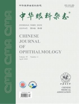
Volume 59 · Issue 04 · 2023
Multilingual International Publishing Platform for Humanities and Social Sciences111
This issue focuses on the research progress in the diagnosis and treatment of ocular surface diseases. The responsible editorial committee responsible for the planning, composition and review of this issue is Professor Yuan Jin.
The advent of artificial intelligence (AI) technology has led to revolutionary advancements in the diagnosis and treatment of ophthalmic diseases, introducing a novel AI-assisted diagnostic approach for ophthalmology that is rich in imaging diagnostic technologies. However, as clinical applications continue to evolve, AI research in ophthalmology faces challenges such as the lack of standardized datasets and innovative algorithm models, insufficient cross-modal information fusion, and limited clinical interpretability. In response to the growing demand for AI research in ophthalmology, it is essential to establish ophthalmic data standards and sharing platforms, innovate core algorithms, and develop clinical logic interpretable models for the screening, diagnosis, and prediction of eye diseases. Additionally, the deep integration of cutting-edge technologies such as 5G, virtual reality, and surgical robots would advance the development of ophthalmic intelligent medicine into a new phase.
Drug-induced keratopathy occurs when the use of certain drugs, particularly ophthalmic preparations, causes pathological changes in the cornea. These changes can be related to the toxic effects of the drugs themselves or drug preservatives. The disease is characterized by a range of clinical features and lacks specific diagnostic criteria, which can lead to misdiagnosis and inappropriate treatment. In order to address these challenges, the Cornea Group of the Ophthalmology Branch of the Chinese Medical Association has brought together leading experts in the field to review key techniques for the diagnosis and treatment of drug-induced keratopathy. As a result, they have developed this consensus to guide the prevention and treatment of this condition.
Meibomian gland dysfunction (MGD) is a common clinical ocular surface disease. In recent years, great progress has been made in basic and clinical research on MGD, and new diagnostic and therapeutic methods have been continuously applied to clinical practice. To improve the understanding of MGD among Chinese ophthalmologists and standardize the diagnosis and treatment of MGD, the Chinese Branch of the Asia Dry Eye Society and relevant academic groups organized experts to discuss the definition and classification of MGD based on recent research progress and clinical experience at home and abroad, forming consensus opinions for the reference of clinicians.
To evaluate the efficiency and safety of collar-button type keratoprosthesis (c-bKPro) implantation for corneal blindness in high-risk transplantation in China.
It was a case series study. High-risk corneal blind patients who planned to undergo c-bKPro implantation were prospectively and continuously enrolled in the Eye Hospital of Shandong First Medical University, Ophthalmology Division of Chinese PLA General Hospital, Zhongshan Ophthalmic Center, Department of Ophthalmology in Eye & ENT Hospital of Fudan University, and Eye Hospital of Wenzhou Medical University from July 2019 to January 2020. The cure for blindness and surgical success were assessed based on visual acuity (VA)≥0.05. The complications and keratoprosthesis retention rate were recorded to determine the safety of the surgery.
Thirty-seven subjects (eyes) were included, of which 32 were male and 5 were female, aged 27 to 72 years old. The indications of c-bKPro implantation were corneal graft failure (21 eyes, 56.8%), chemical injury (8 eyes, 21.6%), thermal burn (5 eyes, 13.5%), unexplained corneal opacity (2 eyes, 5.4%), and corneal perforation (1 eye, 2.7%). Two patients withdrew from the clinical trial at 3 months postoperatively. Thirty-five patients were followed up for 6 months, and 31 were followed up for 12 months. The VA was ≥0.05 in 83.8% of eyes at 6 months and in 81.8% of eyes at 12 months. Among the 11 eyes diagnosed with concurrent glaucoma, 6 eyes achieved a VA of ≥0.05. At 12 months, the c-bKPro retention rate was 100%. The surgical complications included retroprosthetic membrane formation (5 eyes, 16.1%), persistent corneal epithelial defects (5 eyes, 16.1%), macular edema (4 eyes, 12.9%), new-onset glaucoma (4 eyes, 12.5%; including one eye withdrawn from the study at 3 months), sterile corneal melting (2 eyes, 6.5%), sterile vitritis (1 eye, 3.2%), and infectious keratitis (1 eye, 3.2%).
C-bKPro implantation is an effective and safe option for treating corneal blindness in high-risk transplantation in China. Improved visual outcomes could be achieved in most cases, with a relatively low incidence of postoperative complications.
To compare the point-of-care assays for tear matrix metalloproteinase 9 (MMP-9) using domestic and InflammaDry kits, and to evaluate the feasibility of diagnosing dry eye with the domestic kit.
It was a cross-sectional study. Thirty dry eye patients and 30 age-and sex-matched normal volunteers were continuously enrolled in this cross-sectional study from June 2022 to July 2022. Both domestic and InflammaDry kits were used to detect the tear MMP-9 levels. The positive rates were recorded for qualitative analysis, and the gray ratios of bands (the gray value of detection bands to that of control bands) were collected for quantitative analysis. The correlations of MMP-9 levels with age, ocular surface disease index, fluorescence tear break-up time, tear meniscus height, Schirmer′s Ⅰ test score, corneal fluorescein staining score, and meibomian gland dropout were analyzed. The Mann-Whitney U test, paired Chi-square test, Kappa test, and Spearman′s correlation coefficient were used for statistical analysis.
There were 14 males and 16 females (30 eyes) in the control group, and their age was (39.37±19.55) years. In the dry eye group, 11 males and 19 females (30 eyes), aged (46.87±17.85) years, had moderate to severe dry eye. The positive rates of MMP-9 in tear fluid were significantly different between dry eye patients (InflammaDry: 86.67%; domestic kit: 70.00%) and controls (InflammaDry: 16.67%, P<0.001; domestic kit: 6.67%, P<0.001). Although the sensitivity of the domestic kit was lower than that of the InflammaDry kit (70.0% vs. 86.7%, P=0.001), the specificity was higher (93.3% vs. 83.3%, P=0.001). In dry eye patients, the positive coincidence rate was 80.7% (21/26), the negative coincidence rate was 100% (4/4), and the total coincidence rate was 83.3% (25/30), with no significant difference between the two kits (McNemar test: χ2=3.20, P>0.05), and the results of both kits were consistent (Kappa=0.53, P=0.001). The Spearman′s correlation coefficient showed the gray ratios using both kits were positively correlated with the corneal fluorescein staining score (InflammaDry: ρ=0.48, P<0.05; domestic kit: ρ=0.52, P=0.003).
The performances of the domestic and InflammaDry kits are consistent in the point-of-care assay for tear MMP-9, and the domestic kit has lower sensitivity but higher specificity.
To analyze the clinical manifestations and imaging characteristics of in vivo confocal microscopy (IVCM) for Nocardia keratitis.
It was a retrospective case series study. Medical records of 16 consecutive patients (16 eyes) with Nocardia keratitis were collected from the Department of Ophthalmology at Beijing Tongren Hospital, Capital Medical University between 2018 and 2022. The group consisted of 11 males and 5 females. The inclusion criteria for the study were the presence of typical clinical manifestations of Nocardia keratitis and at least one positive pathogenic test (corneal scraping or microbial culture) indicating Nocardia infection. The medical history, clinical and microbiological examination data of the patients were analyzed, including risk factors, diagnosis time, clinical manifestations, diagnostic methods, strain isolation, cure time, and best corrected visual acuity before and after treatment. This study utilized techniques such as slit lamp microscopy, in vivo confocal microscopy (IVCM), scraping cytology, microbial culture, and mass spectrometry identification.
The main risk factors for Nocardia keratitis included plant or foreign body injuries (5 out of 16 cases), contact lens use (4 out of 16 cases), and surgery (2 out of 16 cases). The average time to diagnosis was (20.8±11.8) days, with the shortest time being 8 days and the longest being 60 days. The best corrected visual acuity was less than 0.05 in 7 patients, between 0.05 to 0.3 in 7 patients, and greater than or equal to 0.3 in 2 patients. The typical symptoms included superficial gray-white infiltration in a wreath-like pattern on the cornea, corneal ulcers with dry and gray-white necrotic tissue coverage, and in severe cases, corneal ulcer perforation. Nocardia corneal infection was identified in 12 out of 16 cases by scraping cytology, 9 out of 16 cases by mass spectrometry, and 8 out of 16 cases by both methods. IVCM showed the presence of fine and moderately reflective filamentous hyphae in the subepithelial and superficial stromal layer of the cornea, arranged in elongated, beaded, and branched structures. Infiltration of many hyper-reflective round inflammatory cells was also seen around the hyphae. Fourteen cases were treated with medication and 2 cases were treated with corneal transplantation. The average cure time was (37.5±25.2) days and there were no cases of recurrence during the follow-up period (all greater than 6 months).
Nocardia keratitis is primarily characterized by dense, round, or wreath-like infiltration in the early stage, and by gray-white dry necrotic secretion and hypopyon on the surface of corneal ulcers in the middle and late stages. Fine, branched or beaded, and moderately reflective filamentous structures are the hallmark of the corneal lesion on the IVCM images.
To evaluate the initial safety and efficacy of femtosecond laser-assisted minimally invasive lamellar keratoplasty (FL-MILK) for advanced keratoconus.
It was a case series study. Patients with advanced keratoconus who underwent FL-MILK between August 2017 and April 2020 at Shandong Eye Hospital were prospectively included. The femtosecond laser was used to create an intrastromal pocket in the recipient cornea and a lamellar cornea in the donor. The lamellar cornea was then gently inserted into the intrastromal pocket through the incision and flattened. Clinical measurements included best-corrected visual acuity, 3-mm anterior corneal mean keratometry, anterior and posterior central corneal elevation, central corneal thickness, corneal biomechanics, and endothelial cell density. The follow-up was conducted at 1 month, 12 months, and 24 months after the operation.
There were 33 patients (35 eyes) in the study. Twenty-six patients were male and 7 patients were female. The mean age was (20.34±5.24) years old. All patients completed 12 months of follow-up, and 25 patients (27 eyes) completed 24 months of follow-up. No epithelial ingrowth, infection, or allogeneic rejection was observed. Compared with the preoperative data, the anterior central corneal elevation significantly decreased (P<0.001), the mean keratometry significantly decreased (P<0.05), and the central corneal thickness significantly increased (P<0.001). The corneal biomechanical strength was significantly improved, with the deformation amplitude ratio at 2.0 mm decreasing from 1.39±0.14 preoperatively to 1.21±0.10 at 24 months postoperatively (P<0.001) and the stiffness parameter at the first applanation increasing from 41.49±11.47 preoperatively to 88.41±18.17 at 24 months postoperatively (P<0.001). There were no significant changes in the mean best-corrected visual acuity, posterior corneal elevation, mean spherical equivalent, and endothelial cell density (all P>0.05).
FL-MILK may be a feasible option for advanced keratoconus. This procedure may provide a new resolution for keratoconus.
To investigate the clinical features of dry eye disease in patients with graft-versus-host disease (GVHD) and to identify the correlative factors that contribute to its severity.
It was a retrospective case series study. A total of 62 patients with dry eye disease caused by GVHD after allogeneic hematopoietic stem cell transplantation (HSCT) were recruited from the First Affiliated Hospital of Soochow University between 2012 and 2020. The study population comprised 38 males (61%) and 24 females (39%), with an average age of (35.29±11.75) years. Only the right eye of each patient was evaluated. The patients were divided into two groups based on the severity of corneal epitheliopathy: a mild group (15 eyes) and a severe group (47 eyes). Demographic information, including gender, age, primary disease, type of allogeneic HSCT, donor-to-recipient information, source of hematopoietic stem cells, systemic GVHD, and the time from HSCT to the first visit, was collected. Ophthalmologic assessments, including the Schirmer Ⅰ test, tear breakup time, corneal epithelial staining, and eye margin assessment, were performed during the first visit to the ophthalmology department and compared between the two groups.
The average time from HSCT to the first visit to the ophthalmology department among the 62 patients was (20.26±13.09) months. The median corneal fluorescein staining score was 4.5 points. In the mild group, the main characteristic of corneal staining was scattered punctate staining in the peripheral region in 80% of cases, while in the severe group, corneal staining fused into clumps in both the peripheral region (64%) and the pupillary zone (28%). Results of the Schirmer Ⅰ test were significantly lower in the severe group compared to the mild group (P<0.05). The median total eyelid margin score in the severe group was higher than that in the mild group [9 (7, 12) points vs. 6 (5, 8) points] (P<0.05). The median eyelid congestion score in the severe group was, also higher than that in the mild group [2 (1, 3) points vs. 1 (0, 2) points] (P<0.05). The compatibility between the blood types of the donor and recipient was found to be statistically significant (P<0.05). There was no significant difference in gender, age, family relationship, human leukocyte antigen matching, gender consistency, source of hematopoietic stem cells, or the occurrence of systemic GVHD between the two groups (P>0.05).
Patients in the mild group had scattered punctate corneal staining in the peripheral region, while those in the severe group showed fusion of corneal staining into clumps in both the peripheral and pupillary zones. The severity of dry eye disease caused by GVHD was strongly correlated with eyelid margin lesions. A higher degree of eyelid margin lesions indicated more severe dry eye disease caused by GVHD. Additionally, compatibility between the blood types of the donor and recipient may play a role in the development of GVHD-associated dry eye.
This 15-year-old male patient has been diagnosed with osteogenesis imperfecta through genetic testing after birth and has poor vision. His full corneas in both eyes are unevenly thinned and bulging in a spherical shape, with the right eye being more severe. He underwent a limbal stem cell-sparing lamellar keratoplasty in the right eye, resulting in improved vision with a corrected visual acuity of 0.5, a decrease in corneal curvature, and a significant increase in corneal thickness. The surgery had a satisfactory outcome. The condition of the left eye is progressing and will require further surgical treatment.
This article presents a case of orbital rhabdomyosarcoma (RMS) in a child who also had neurofibromatosis type 1 (NF-1). Although NF-1 is a common neurogenetic disease, there are few reports of its combination with orbital RMS. The patient underwent surgical removal of the tumor at the age of one, but the cancer recurred after five years. Following pathological and genetic diagnosis, the patient was confirmed to have orbital RMS combined with NF-1. After surgical treatment and chemotherapy, the patient′s eye condition has stabilized. This article focuses on analyzing the clinical manifestations of this case and reviews relevant literature to improve our understanding of this disease in children.
A 63-year-old male with a healthy history presented with a red and swollen right eye for 3 months. Neuro-ophthalmic examination showed slight bulging of the right eyeball, and multiple spiral conjunctival vessels were visible on the surface of the right conjunctiva, suggesting a right carotid cavernous fistula. Cerebral angiography showed left occipital dural arteriovenous fistulas. After endovascular embolization treatment, the patient′s abnormal craniocerebral venous drainage and right eye syndrome resolved, and there was no recurrence during the one-month clinical follow-up after surgery.
The patient was a 53-year-old male. He went to the Department of Ophthalmology of the Second Hospital of Hebei Medical University because of the dark shadow in front of his right eye fluttering for 1 month and visual acuity decreasing with visual object occlusion for 20 days. Eye examination: visual acuity was 0.15 for the right eye and 1.0 for the left eye; No obvious abnormalities were observed in the anterior segment of both eyes; Retinal detachment can be seen above the right fundus, and 2 horseshoe-shaped hiatus can be seen above the detached retina (Quintessential Picture 1). Clinical diagnosis: Rhegmatogenous retinal detachment in the right eye.
The patient was a 28-year-old male. He went to Jinan Mingshui Eye Hospital because of repeated redness of his left eye and tearing for 1 year. Eye examination: visual acuity was 1.2 in the right eye and 1.0 in the left eye; IOP was 15 mmHg (1 mmHg =0.133 kPa) in the right eye and 18 mmHg in the left eye; A granuloma was seen in the conjunctiva of the upper eyelid of the left eye, and an insect-like foreign body was seen in the central area of the granuloma, with white substances attached around the foreign body, surface telangiectasia, conjunctiva hyperemia, corneal epithelial defects, and no obvious abnormalities were found in other ocular structures (Quintessential Picture 1). Clinical diagnosis: conjunctival granuloma of upper eyelid of left eye; Insect foreign body in the conjunctiva of the upper eyelid of the left eye.
The patient was a 24-year-old female. Because of grinding pain in both eyes and blurred vision for 5 days, he went to Hebei Eye Hospital. He had been myopic in both eyes for 12 years and wore contact lenses intermittently. Eye examination: visual acuity was 0.12 for the right eye and 0.1 for the left eye; IOP was 15.0 mmHg (1 mmHg =0.133 kPa) in the right eye and 14.0 mmHg in the left eye; The conjunctiva of the right eye was mixed congested, with an inverted triangular epithelial defect of about 2 mm ×3 mm in the central cornea, mild edema of the corneal epithelium in other parts, vague intraepithelial microvesicle-like changes, and no edema of the corneal stroma; The conjunctiva of the left eye is mildly congested, the corneal epithelium is rough, and a large number of microvesicle-like changes with basically the same shape and size can be seen in the corneal epithelium in the central area, with a range of about 6 mm ×7 mm. The corneal fluorescein staining is positive, the corneal epithelium in other parts is smooth, and there is no edema in the corneal stroma (Essence Picture 1); The anterior chamber of both eyes was moderately deep, the aqueous humor was clear, the pupil was round, about 3 mm in diameter, and the response to light was normal. Clinical diagnosis: binocular Meesmann corneal dystrophy.
Neurotrophic corneal disease is a degenerative eye condition that occurs due to damage to the trigeminal nerve. This condition presents as a persistent corneal epithelial defect, corneal ulceration, or even perforation, and the main cause is a loss of corneal nerve function. While traditional treatments mainly focus on supportive measures to repair corneal damage, they cannot cure the condition completely. A new surgical treatment option called corneal sensory reconstruction surgery can rebuild the corneal nerve, slow down the progression of the corneal disease, promote corneal epithelial repair, and improve vision. This article reviews the surgical techniques used in corneal sensory reconstruction, including direct nerve repositioning and indirect nerve transplantation, and discusses their treatment outcomes and future prospects.
The prevalence of dry eye in children is increasing with changes in the environment and the widespread use of electronic products. However, due to poor ability to express themselves and hidden symptoms of children, lack of understanding of dry eye in children, children with dry eye are likely to be misdiagnosed. Dry eye can seriously affect the quality of children′s learning, life, vision and visual development. Therefore, it is urgent to raise awareness of clinical workers about dry eye in children, prevent the occurrence of related complications of dry eye, and avoid permanent visual damage to children. This review discusses and summarizes the epidemiology and common risk factors of children with dry eye, with the aim of improving doctors′ understanding of dry eye in children.
Monkeypox is the most important orthopoxvirus infectious disease in humans after the extinction of smallpox. Recent outbreaks of monkeypox in several countries show obvious human-to-human transmission, which has aroused great concern around the world. Monkeypox infection can also affect the eyes. This article reviews the clinical features and ocular manifestations of monkeypox virus infection in order to attract the attention of ophthalmologists.
Viral endotheliitis is a common cause of corneal endothelial decompensation, and its clinical manifestations are diverse and complex, which can easily lead to misdiagnosis and misdiagnosis. Based on the "Chinese Expert Consensus on the Diagnosis and Treatment of Viral Keratoendotheliitis (2023)", this article elaborates on the classification, clinical characteristics, imaging diagnosis, molecular biology and immunological diagnostic points, differential diagnosis and treatment plan of viral keratoendotheliitis, in order to provide reference for clinical work.
CURRENT ISSUE