Multilingual International Publishing Platform for Humanities and Social Sciences111
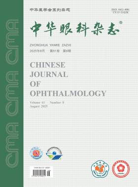
Volume 61 · Issue 08 · 2025
Multilingual International Publishing Platform for Humanities and Social Sciences111
The key report content of this issue is ocular surface diseases. Professor Xu Jianjiang is the responsible editorial board member in charge of the planning, composition and review of this issue.
By summarizing the diverse pathogens (viruses, bacteria, fungi, amoebae) and clinical features of infectious corneal epitheliopathy, this editorial clarifies the key points in diagnosis and differential diagnosis. Emphasizing the golden window of epithelial-stage intervention, it advocates a comprehensive diagnostic approach integrating medical history, slit-lamp findings, microbiological tests (corneal scraping, special staining/culture), in vivo confocal microscopy, and molecular diagnostics (tear virus antibodies, metagenomic sequencing). The tripartite evidence chain of "history-imaging-pathogen" is proposed to transcend morphology-dependent empiricism, with strict caution against glucocorticoid use before etiological confirmation. Standardized precision diagnosis at the epithelial stage is pivotal for early intervention and visual prognosis improvement.
In recent years, cases of microsporidial keratoconjunctivitis in China have been increasingly reported. Patients are often misdiagnosed as viral or fungal keratitis before confirmation, leading to prolonged disease course due to inappropriate treatment, and clinical awareness of this disease has long been insufficient. Current diagnosis and treatment face multiple challenges: limited understanding of the biological characteristics of microsporidia and risk factors such as parrot exposure, difficulty in clinical differentiation between keratoconjunctivitis and stromal keratitis types, and the absence of uniform diagnostic and therapeutic standards. It is imperative to strengthen clinical awareness of early diagnosis for patients with risk factors, promptly launch multicenter studies to establish standardized diagnostic and therapeutic consensus, so as to improve overall management and ensure patients receive timely and effective treatment.
Keratoconus is the most common blinding eye disease among young adults. The treatment methods comprise non-surgical interventions (in the early stage) and surgical interventions (in the advanced stage). Surgical approaches mainly include minimally invasive procedures such as corneal collagen cross-linking and minimally invasive lamellar keratoplasty, as well as more invasive procedures like lamellar or deep anterior lamellar keratoplasty and penetrating keratoplasty. As keratoconus progresses, the optimal timing and selection of surgical methods warrant focused attention and discussion. This article proposes three guiding principles on the selection of minimally invasive surgical approaches and the timing for keratoconus: 1) minimizing surgical trauma, 2) enhancing procedural precision, and 3) appropriately advancing the timing of surgical intervention. Through in-depth discussions, these principles aim to provide valuable references and clinical guidance for ophthalmic practice.
With the emergence of various multi-segment myopic defocus design spectacles, evaluating and verifying their myopia control effectiveness and controlling their quality and wearing safety have gradually attracted widespread attention. At present, myopia prevention and control spectacles have not been classified as medical devices directly supervised by relevant national departments in China. Multi-segment myopic defocus design spectacles, as an important intervention for retarding the progression of myopia, especially for children and adolescents, should ensure the myopia control effectiveness, safety, and adherence to standardized fitting protocols. Aiming to provide guidance and reference for healthcare institutions and practitioners, the Society of Contact Lens Safety Monitoring & Vision Health of Chinese Health Association and the Western Optometry Expert Committee of Sichuan Optometry Society, based on the current evidence-based medical research, combined with practical experience and extensive solicitation of opinions, have reached consensus opinions on the quality control, effectiveness evaluation (including long-term effectiveness), and safety evaluation of multi-segment myopic defocus design spectacles, as well as the service norms for the fitting of spectacles.
To investigate the clinical and etiological characteristics of microsporidial keratitis (MK).
A retrospective case series study was conducted. Clinical data were collected from patients diagnosed with MK at Beijing Tongren Hospital, Capital Medical University between June 2023 and April 2025, including risk factors, time to diagnosis, clinical manifestations, laboratory findings, best-corrected visual acuity (BCVA) before and after treatment, and cure duration. In addition to routine microbiological smear cytology, specimens underwent in vitro Vero cell culture for isolation and identification. Polymerase chain reaction (PCR) targeting the small subunit ribosomal RNA gene of microsporidia was performed on corneal lesion secretions and parrot feces (suspected infection sources), followed by sequencing and strain identification to clarify the infection origin.
Ten patients (12 affected eyes) were enrolled, comprising 4 males and 6 females with a median age of 36 years (range: 7 to 77 years). Major risk factors included parrot contact (8/10), plant-induced trauma (1/10), and soil-related trauma (1/10). The median interval from symptom onset to diagnosis was 12.5 days (range: 3 to 56 days). At presentation, 2 eyes had BCVA<0.5, and 10 eyes had BCVA≥0.5. Typical manifestations included conjunctival follicular reaction and scattered multifocal punctate elevated epithelial lesions on the corneal surface with positive fluorescein staining. Stromal involvement presented as pseudo-dendritic epithelial defects, diffuse stromal infiltration/edema, neovascularization, and positive keratic precipitates. Microsporidial spores were detected in all 12 eyes via scraping cytology. Vero cell culture was positive in 4 eyes (33.3%), and all PCR assays were positive. In vivo confocal microscopy revealed scattered hyper-reflective puncta in the epithelium, central erosive lesions in epithelial cells, and rose-like aggregates of damaged epithelial cells. Densely arranged punctate structures were observed in the stroma upon stromal involvement. All 9 epithelial MK patients achieved clinical cure after pharmacotherapy, with a median treatment duration of 21 days (range: 10 to 40 days).
MK presents as epithelial or stromal type, with the former having a favorable prognosis. Parrot contact and ocular trauma are key risk factors. Vero cells are suitable for microsporidial isolation/culture. The diagnosis relies on typical clinical signs, corneal scraping microbiology, and PCR analysis.
To investigate the distribution patterns, clinicopathological characteristics, and age-specific trends of conjunctival tumors in a single center from June 2018 to December 2023, and to analyze their changes compared with historical data.
This was a retrospective case series study. A total of 1 413 histopathological cases of excised conjunctival lesions from the Second Affiliated Hospital of Zhejiang University School of Medicine between June 2018 and December 2023 were enrolled as the study cohort. All cases underwent histopathological classification, with partial specimens supplemented by immunohistochemical staining, special staining, or electron microscopy. The histopathological types (epithelial, melanocytic, inflammatory lesions, etc.), lesion nature (benign, premalignant, or malignant), and age distribution (children and adolescents, ≤18 years; young-middle-aged adults, 19 to 59 years; the elderly, ≥60 years) of these cases were compared with those of 1 233 historical cases (January 2000 to May 2018).
Among 1 216 eligible cases, there were 567 males and 649 females, with 1 052 benign lesions (86.51%) and 164 premalignant/malignant lesions (13.49%). Histopathologically, epithelial tumors (508 cases, 41.86%) and melanocytic tumors (433 cases, 35.59%) were predominant, followed by inflammatory lesions (118 cases, 9.70%), lymphoproliferative lesions (59 cases, 4.85%), and vascular tumors (55 cases, 4.52%). Benign lesions were dominated by cysts (323 cases, 26.56%) and nevi (314 cases, 25.82), while premalignant/malignant lesions were primarily conjunctival intraepithelial neoplasia (68 cases, 5.59%) and lymphoma (40 cases, 3.29%). Compared with historical data, the study cohort showed a significantly higher proportion of benign lesions (86.51% vs. 78.48%) and a lower proportion of premalignant/malignant lesions (13.49% vs. 21.52%). The proportion of cysts increased from 12.89% to 26.56% (becoming the most common benign lesion), while squamous papilloma decreased from 10.52% to 6.74%. In age distribution, young-middle-aged adults accounted for 62.83% (764 cases) in the study cohort (slightly higher than 60.00% in historical cases), and the elderly accounted for 23.19% (282 cases) (slightly lower than 24.27%).
Analysis of single-center data from 2018 to 2023 revealed an increased proportion of benign conjunctival lesions and a decreased proportion of premalignant/malignant lesions, with epithelial and melanocytic tumors remaining predominant but inflammatory lesions decreasing. Children, adolescents, and young-middle-aged adults primarily presented with cystic and nevus-like benign lesions, while the elderly, also showing benign lesions, had a significantly higher proportion of premalignant/malignant lesions.
To investigate the changes in ocular surface damage signs in patients with non-small cell lung cancer (NSCLC) after treatment with rearrangement during transfection (RET) inhibitors.
This was a retrospective case series study. Clinical data were collected from 7 NSCLC patients (14 eyes) who developed dry eye symptoms after receiving RET inhibitor chemotherapy at the Second Affiliated Hospital of Zunyi Medical University from October 2023 to December 2024. All patients presented to the ophthalmology department due to dry eye symptoms occurring at different time points after RET inhibitor treatment. Corneal fluorescein staining scores, tear film breakup time (BUT), and Schirmer Ⅱ test results were recorded at different time periods post-treatment. Patients were treated with artificial tears, meibomian gland massage, and eyelid heat compress, and some patients were additionally prescribed cyclosporine A eye drops. Follow-up was conducted to observe the improvement of dry eye symptoms and ocular surface damage.
A total of 7 patients were included, consisting of 5 males and 2 females, with an average age of (50.6±5.0) years. The time from initiation of treatment to the onset of ocular symptoms was (87.7±54.3) days. All patients had corneal fluorescein staining scores>1, BUT<5 seconds, and Schirmer Ⅱ test results<5 mm. No abnormalities were detected in corneal endothelial cell count, macular optical coherence tomography, ultra-wide field fundus imaging, or fundus autofluorescence. During the treatment period, 3 patients (42.9%) developed simultaneous peeling of palm skin and nails. After treatment, symptoms improved in 4 patients, and the remaining 3 patients showed improvement after adding cyclosporine A eye drops. One patient discontinued the RET inhibitor due to severe dry eye symptoms, with subsequent improvement; however, symptoms recurred and worsened upon resumption of the medication.
NSCLC patients receiving RET inhibitor treatment may develop ocular surface damage characterized by corneal epithelial injury, reduced tear film stability, and decreased tear secretion. Clinicians should pay close attention to the ocular adverse reactions and associated mucocutaneous manifestations of these drugs.
To investigate the effect of dimethyl itaconate (DMI), an NRF2 activator, on hypoxia-induced scleral fibroblasts.
It was a experimental study. A guinea pig model of form-deprivation myopia (FDM) was established. Ocular refraction and axial length were measured using eccentric infrared photorefraction and A-scan ultrasonography, respectively. Western blot was used to detect NRF2 protein expression in the sclera of FDM guinea pigs. Primary guinea pig scleral fibroblasts (GSFs) were cultured and identified via immunofluorescence staining for vimentin and cytokeratin. GSFs at passages 3-5 were used for experiments. For the hypoxic treatment, GSFs were cultured in a hypoxia incubator chamber with 1% O₂, 5% CO₂, and 94% N₂, with cells cultured under 21% O₂ serving as the normoxia control. After 48 h of culture, Western blot analysis was performed to detect the expression of HIF-1A, COL1A1, MMP2, and NRF2 in GSFs. The mRNA expression of the NRF2 downstream target gene NQO1 mRNA was detected by quantitative real-time polymerase chain reaction (qRT-PCR). Nuclear translocation of NRF2 was assessed by immunofluorescence staining. For the DMI treatment, GSFs were treated with 150 μmol/L DMI or an equal volume of PBS for 3 h, followed by the hypoxic treatment for 48 h. Subsequent analyses (Western blot, qRT-PCR, and immunofluorescence staining) were performed as described above.
After 4 weeks of form deprivation, guinea pigs in the FDM group exhibited a significant myopic shift in refraction [(-3.248±1.501) vs. (4.143±0.350)D; P<0.001] and increased axial length [(8.650±0.081) vs. (8.363±0.020) mm; P<0.05] compared to the normal control group. NRF2 protein expression in the sclera of FDM guinea pigs was significantly decreased (0.347±0.110 vs. 0.796±0.032; P<0.05). GSFs migrated from scleral tissue pieces 7-9 days after culture and reached confluence after approximately 14 days. At low density, GSFs were stellate-shaped; after confluence, they organized into bundle-or swirl-shaped patterns. Immunofluorescence results confirmed GSFs were positive for vimentin and negative for cytokeratin. Compared with the normoxia group, the hypoxia group showed increased protein expression of HIF-1A (1.361±0.043 vs. 0.843±0.053; P<0.001) and MMP2 (0.640±0.042 vs. 0.355±0.052; P<0.05), but decreased COL1A1 (0.451±0.063 vs. 0.701±0.043; P<0.05). NRF2 protein (0.154±0.023 vs. 0.399±0.026, P<0.05) and NQO1 mRNA (0.700±0.050 vs. 1.000±0.004, P<0.05) expression was reduced, and the NRF2 nuclear translocation was diminished in the hypoxia group. Compared with the PBS group, the DMI-treated group showed increased NRF2 protein (0.361±0.056 vs. 0.146±0.023; P<0.05) and NQO1 mRNA (1.775±0.028 vs. 1.001±0.022; P<0.001) expression, enhanced NRF2 nuclear translocation, increased COL1A1 protein (0.842±0.082 vs. 0.400±0.054; P<0.05), and decreased MMP2 protein (0.547±0.077 vs. 0.841±0.039; P<0.05).
Activation of NRF2 participates in scleral remodeling in myopia by promoting COL1A1 expression and inhibiting MMP2 expression in scleral fibroblasts, suggesting NRF2 as a potential therapeutic target for myopia.
To investigate the accuracy of the gaze-tilt test in predicting the postoperative head posture improvement in patients with congenital superior oblique palsy (CSOP).
A diagnostic test was conducted, enrolling children aged 3 to 6 years diagnosed with unilateral CSOP and scheduled for unilateral inferior oblique weakening surgery at Tianjin Eye Hospital between May 2023 and January 2024. Preoperatively, the single eye cover test and the gaze-tilt test were used to assess the patients′ compensatory head posture improvement. The overall predictive accuracy, predictive accuracy for different head position improvement outcomes, and error rates (false positive rate and false negative rate) of the two methods were compared by evaluating the predicted outcomes against the actual head postures at postoperative day 1 and month 1. The paired t-test, paired Wilcoxon signed-rank test, and paired chi-square test were used for statistical analysis.
Thirty-two children were included in the final analysis, consisting of 17 males and 15 females. The preoperative head tilt angle was 77.50°(75.00°, 80.75°). The head tilt angles at postoperative day 1 and month 1 were 88.00°(82.64°, 89.99°) and 88.90°(87.35°, 90.00°), respectively. The gaze-tilt test predicted the postoperative head position tilt angle of 87.00°(86.00°, 89.07°), which was significantly different from the angle of 83.00°(80.27°, 87.30°) predicted by the single eye cover test (P<0.05). Compared with the actual tilt position angle on postoperative day 1, the gaze-tilt test showed no statistically significant difference (P>0.05), but the single eye cover test showed significant difference (P<0.05). The predictive accuracy of the complete head position improvement was significantly higher for the gaze-tilt test [postoperative day 1: 86.36%(19/22), postoperative month 1: 81.48% (22/27)] compared to the single eye cover test [postoperative day 1: 50.00% (11/22), postoperative month 1: 44.44% (12/27)] (both P<0.05). The false negative rate of the gaze-tilt test was lower than that of the single eye cover test. The prediction error of the gaze-tilt test (postoperative day 1:-0.53°±4.17°, postoperative month 1: 1.67°±4.26°) was smaller than that of the single eye cover test (postoperative day 1: 2.81°±4.61°, postoperative month 1: 5.01°±4.90°), with statistically significant differences (both P<0.05).
Compared to the single eye cover test, the gaze-tilt test demonstrated higher accuracy and smaller predictive errors in predicting the postoperative head posture improvement in patients with CSOP.
A 9-year-old male patient with binocular vision loss for 3 months visited the Department of Ophthalmology of the Jalaid Banner Mongolian Medical General Hospital. The optic disc in both eyes was ruddy and moderately edematous with a blurred margin, and the disc periphery was markedly edematous and elevated. Ocular B-scan ultrasonography and optical coherence tomography (OCT) examinations showed varying degrees of optic disc elevation. Enhanced depth imaging OCT examination revealed a slightly hyper-reflective ovoid mass-like structure with clear boundaries laterally protruding under the retinal nerve fiber layer and above the Bruch′s membrane at the site corresponding to the optic disc elevation. The 3D-reconstructed OCT image of the optic disc showed the optic disc elevation and the slightly hyper-reflective ovoid mass-like structure. The clinical diagnosis was pseudopapilledema combined with a peripapillary hyper-reflective ovoid mass-like structure in both eyes. Finally, the patient was followed up in the ophthalmology outpatient department.
The patient was a 68-year-old male. He went to Hebei Eye Hospital due to redness and pain in his right eye with visual acuity loss for 2 and a half months. Previous old cerebral infarction 2 years. Eye examination: visual acuity is manual in the right eye and 1.0 in the left eye; IOP was 15.3 mmHg (1 mmHg =0.133 kPa) in the right eye and 16.6 mmHg in the left eye; Moderate mixed congestion of the conjunctiva of the right eye, a small amount of secretion can be seen in the conjunctival sac, a grayish-white infiltration focus of about 6 mm ×6 mm in the center of the cornea, and a tan-brown mycelial coat can be seen attached to the surface of the lesion. Other parts of the cornea are transparent, the depth of the anterior chamber is normal, and no empyema is seen (A in essence picture 1). The scraping of the right corneal lesion showed tan-brown branched hyphae under light microscope by Giemsa staining (B in Quintessential Picture 1), and branched hyphae structure by fluorescence staining (C in Quintessential Picture 1). Clinical diagnosis: pigmented fungal keratitis of the right eye.
The patient was a 15-year-old male. Because of blurred vision in both eyes, he went to the Department of Ophthalmology, the Second Affiliated Hospital of Army Medical University. He was diagnosed with right sensorineural deafness in our hospital 3 years ago; The mother had a history of similar disease with heterochromatic iris in both eyes. Eye examination: the naked eye visual acuity was 0.15 in the right eye, 0.25 in the left eye, and the corrected visual acuity of both eyes was 1.0; See essence picture 1 for the anterior segment of both eyes, and essence picture 2 for the fundus. Clinical diagnosis: Waardenburg syndrome; Refractive error in both eyes.
Dry eye, a clinically prevalent chronic ocular surface disorder, poses significant therapeutic challenges for moderate-to-severe patients due to abnormal tear secretion and ocular surface microenvironment imbalance. This article presents a comprehensive review of neuromodulation techniques in dry eye intervention, focusing on non-invasive therapeutic approaches including electrical stimulation, magnetic stimulation, mechanical stimulation, and pharmacological neurostimulation. Specifically, intranasal tear neurostimulation (such as TrueTear) promotes tear secretion by activating the nasolacrimal reflex pathway, while transcutaneous electrical stimulation and transcranial magnetic stimulation improve tear film stability by regulating neural conduction pathways. Mechanical stimulation devices and pharmacological neuroactivators have been confirmed in clinical studies to effectively alleviate both subjective symptoms and objective signs in dry eye patients. Although the therapeutic value of neuromodulation in dry eye has been widely recognized, precise elucidation of its mechanism, evaluation of efficacy differences across dry eye subtypes, and long-term safety considerations still require further investigation. By integrating the latest research findings in the field, this review aims to provide theoretical references and novel clinical perspectives for precision neuromodulation therapy in dry eye management.
Suture plays the role of closing the incision, fixing and shaping the tissue structure in cataract extraction surgery. Different methods and techniques can be used according to different situations to obtain good surgical results. Based on clinical experience, this article expounds and summarizes the application of suture and needle in complex cataract extraction surgery, including fixing posterior chamber intraocular lens and capsular bag tension ring, repairing iris, etc., in order to provide reference and guidance for clinical related work.
CURRENT ISSUE