Multilingual International Publishing Platform for Humanities and Social Sciences111
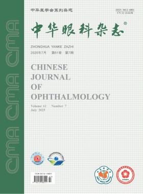
Volume 61 · Issue 07 · 2025
Multilingual International Publishing Platform for Humanities and Social Sciences111
This issue focuses on the research progress of strabismus and pediatric ophthalmology diagnosis and treatment. The responsible editorial committee responsible for the planning, composition and review of this issue is Professor Liu Hu.
The critical period for visual function and ocular structure development occurs from 0 to 6 years of age, making standardized eye care and vision screening services during this stage vital for improving lifelong visual outcomes and quality of life. In China, policy-driven initiatives have established a preliminary eye care and vision screening service system for this age group. However, problems such as insufficient public awareness, limitations in screening technology, fragmented service networks, and decentralized data management systems hinder early intervention and follow-up of abnormal visual development. To address these issues, a collaborative effort involving the government, healthcare institutions, schools, and families is essential. By integrating clinical medicine with preventive care, a tiered service network for medical care should be developed. Leveraging artificial intelligence to optimize screening technologies and establishing a nationally unified, standardized child eye health information management system are crucial to create a closed-loop, digitalized management model integrating health education, screening and early warning, precise diagnosis and treatment, visual rehabilitation, and intelligent follow-up. Multisectoral action is essential to advance pediatric eye care and vision screening services and lay the foundation for lifelong ocular health management.
The World Health Organization proposed the global elimination of avoidable blindness in 2020. Screening for eye diseases in infants and young children serves as a fundamental approach to blindness prevention, holding significant importance. This article highlights the critical role of infant and young child eye disease screening from three perspectives: its position as a key component in the childhood blindness prevention, the current status and deficiencies in China′s screening practices, and the establishment of effective screening models. The importance of eye disease screening in early childhood should be emphasized, which would help to enhance the standards of pediatric eye disease screening and advance blindness prevention efforts in China.
Congenital pupillary membranes, as common developmental anomalies of the anterior segment, primarily include persistent pupillary membrane (PPM) and anterior-anterior type of persistent fetal vasculature (AAPFV). Although both manifest as residual membranous tissue in the pupillary area, they differ significantly in embryonic origin and clinical characteristics. PPM arises from incomplete regression of the embryonic pupillary membrane, presenting as non-progressive filamentous or web-like tissue with iris coloration. In contrast, AAPFV is associated with abnormal regression of the primitive hyaloid vascular system, characterized by progressive white fibrovascular membranes that may lead to secondary glaucoma and deprivation amblyopia, thus necessitating early surgical intervention. This article systematically elucidates the core distinctions between these two pupillary membranes in terms of clinical presentation, disease progression, and management strategies, emphasizing the need for ophthalmologists to improve diagnostic recognition to prevent misdiagnosis and inappropriate management.
Strabismus is a common condition in ophthalmology, and surgical intervention is a primary treatment approach. However, there is currently a lack of uniform standards and guidelines for the perioperative management of pediatric strabismus in China. To further standardize the preoperative and postoperative management, improve surgical outcomes, and ensure patient safety in clinical practice, the Chinese Association for Pediatric Ophthalmology and Strabismus along with the Pediatric Ophthalmology and Strabismus Group of Chinese Ophthalmologist Association has developed consensus opinions on the perioperative management of pediatric strabismus, based on clinical research from both domestic and international sources, comprehensive reviews of the literature, expert clinical insights, and thorough discussions. The consensus provides guidance for the preoperative assessment and surgical preparation, intraoperative and postoperative complication management, and postoperative follow-ups and care in pediatric strabismus.
To investigate the clinical characteristics of the inverted Brown pattern in children and the surgical efficacy of inferior oblique weakening for the disease.
It was a retrospective case series study. The medical records of 11 patients (11 eyes) who were diagnosed with congenital superior oblique palsy with the inverted Brown pattern and underwent strabismus surgery in Tianjin Eye Hospital were reviewed. Their pre-and post-operative motility patterns, vertical deviation angle and fundus torsion, intraoperative forced duction test findings of oblique muscles were analyzed. Statistical analyses were conducted using the t-test, ANOVA, and Tukey′s test.
There were 9 male and 2 female patients, with an average age of 5.5 (3.0, 7.0) years. Four patients previously received oblique muscle surgery for congenital superior oblique palsy. Weakening of the inferior oblique muscle was performed in all patients. The follow-up period was 6 to 8 months. The pre-and post-operative vertical deviation angles in primary gaze were significantly different at 33 cm (11.2Δ±2.8Δvs. 1.1Δ±1.3Δ; t=13.6, P<0.05) and at 5 m (11.1Δ±2.9Δvs. 0.7Δ±1.0Δ; t=12.1, P<0.05). The mean hyperdeviation in down-and-in gaze of the higher eye was 18.1±6.0 preoperatively and 0.8Δ±1.5Δ postoperatively, showing significant difference (t=8.2, P<0.05). The vertical misalignment of the higher eye in up-and-in gaze was 5.3°±4.5° before surgery and 0.3°±0.9° after surgery, with significant difference (t=3.3, P<0.05). There was also a significant difference between the pre-and post-operative fundus torsion angles (ex20.2°±3.5° vs. ex12.8°±4.6°; t=3.2, P<0.05). The torsional forced duction test of inferior oblique measurements yielded internal rotation angles of 14.5°±5.2° and 35.9°±4.4° intraoperatively for the higher eye and the contralateral eye, and 58.6°±7.1° postoperatively for the higher eye, with significant difference (F=165.7, P<0.001).
The deficiency of depression in adduction observed in the inverted Brown pattern is caused by inferior oblique muscle tightness. Weakening of the inferior oblique muscle can effectively correct the misalignment in primary gaze and downward gaze.
To analyze the clinical characteristics, surgical treatment, and prognosis of pediatric retinal detachment associated with Stickler syndrome.
This retrospective case series study included 20 eyes from 18 patients aged ≤18 years diagnosed with Stickler syndrome-related retinal detachment at the Department of Ophthalmology, Beijing Tongren Hospital, Capital Medical University between January 1, 2018 and August 1, 2023. All patients underwent comprehensive ophthalmological evaluations. The personalized retinal reattachment surgery plan was selected based on individual factors, including age, visual acuity, intraocular pressure, anterior segment status, and vitreoretinal conditions. The follow-up duration ranged from 12 to 72 months. Postoperative outcomes, including visual acuity, intraocular pressure, retinal reattachment status, and complications, were assessed.
There were 16 males (17 eyes) and 2 females (3 eyes), with a mean age of 9 (5, 11) years. All 20 eyes (100%) presented with rhegmatogenous retinal detachment, of which 13 eyes (65.0%) exhibited C1-D2 proliferative vitreoretinopathy, 11 eyes (55.0%) had total retinal detachment, 7 eyes (35.0%) showed giant retinal tears, and 6 eyes (30.0%) were complicated by choroidal detachment. Surgical interventions included pars plana vitrectomy (PPV) with silicone oil tamponade in 11 eyes (55.0%), scleral buckling alone in 5 eyes (25.0%), combined PPV with silicone oil tamponade and scleral buckling in 3 eyes (15.0%), and PPV with C3F8 gas tamponade in 1 eye (5.0%). Two eyes (10.0%) experienced recurrent retinal detachment after the initial surgery and required a secondary procedure. Among the 14 eyes treated with silicone oil tamponade, successful oil removal was achieved in 7 eyes (50.0%). At the final follow-up, the median best-corrected visual acuity significantly improved to logMAR 1.00 (0.70, 1.19) from preoperative logMAR 2.0 (1.33, 2.60) (Z=-3.83, P<0.001). The anatomical success rate of retinal reattachment was 100%, with no significant postoperative complications observed in any patient.
Retinal detachment associated with Stickler syndrome predominantly manifests as rhegmatogenous retinal detachment, frequently complicated by severe proliferative vitreoretinopathy, giant retinal tears, and choroidal detachment. Selecting an appropriate surgical approach based on distinct clinical characteristics can effectively achieve retinal reattachment, improve vision function, and promote normal development of the affected eye.
To investigate changes in diffusion-weighted imaging (DWI) of extraocular muscles in patients with intermittent exotropia (IXT) during binocular convergence using MRI, and to explore the imaging characteristics of extraocular muscle alterations in IXT.
It was a cross-sectional study. Patients diagnosed with IXT at the First Affiliated Hospital of Anhui Medical University from January 2023 to September 2023 were enrolled as the IXT group, with age-and sex-matched healthy individuals recruited as the control group. Axial MRI scanning was used to measure DWI signal intensity values of the medial rectus, lateral rectus, and optic nerve during primary gaze and convergence gaze. The normalized DWI values between the two groups and two gaze conditions were compared. The correlation between normalized DWI value changes and the degree of strabismus in the IXT group was analyzed. Normally distributed data were analyzed using independent and paired t-tests, while non-normally distributed data were evaluated with Mann-Whitney and Wilcoxon signed-rank tests. Pearson correlation analysis was used to assess the relationship between DWI values and the degree of strabismus.
Eleven patients (22 eyes) were included in the IXT group (six males, five females; mean age, 24±4 years), and 10 healthy controls (20 eyes) were included (four males, six females; mean age, 24±1 years). Baseline characteristics were comparable between groups (P>0.05). In the control group, the normalized DWI value of the medial rectus was 0.86 (0.72, 0.97) during primary gaze and 0.97 (0.87, 1.20) during convergence gaze, with a significant difference (P<0.05). In the IXT group, the normalized DWI value of the medial rectus was 0.77 (0.72, 0.93) during primary gaze and 0.81 (0.69, 1.08) during convergence gaze, with no significant difference (P>0.05). A negative correlation trend was observed between changes in normalized DWI values of the medial rectus during convergence gaze and the degree of strabismus in the IXT group (r=-0.58, P=0.059).
Patients with IXT exhibit significantly reduced changes in normalized DWI values of the medial rectus during binocular convergence compared to healthy controls.
To investigate the impact of maternal factors during pregnancy and birth factors on astigmatism in children.
It was a case-control study. Patients aged 3 to 12 years old were continuously selected from October 2023 to January 2024 in the Pediatric Eye Care Department of Qinhuangdao Maternal and Child Health Care Hospital. Children with astigmatism were included as the case group, and children without astigmatism were included as the control group. All children underwent cycloplegic autorefraction. Astigmatism was defined as a cylinder refractive error with an absolute value of≥1.00 diopter. Only the right eye were included in the analysis. The maternal factors during pregnancy [maternal age at delivery, delivery mode, paternal age, progesterone during pregnancy, pre-pregnancy body mass index (BMI), gestational weight gain, smoking during pregnancy, hypertensive disorders of pregnancy, gestational diabetes mellitus] and children′s birth history [birth weight, birth order, BMI, preterm birth, nuchal cord] were recorded. The independent t-test, non-parametric test, and Chi-square test were used for comparison between groups. Multiple logistic regression analysis was performed and risk factors during pregnancy for astigmatism in children were identified by calculating the odds ratio (OR) and its 95% confidence interval (95%CI).
A total of 146 children (146 eyes) were included in the case group, consisting of 73 males and 73 females with a median age of 6 (5, 9) years, and 161 children (161 eyes) were included in the control group, consisting of 73 males and 88 females with a median age of 7 (5, 9) years. No statistically significant differences in age and gender were found between the two groups (all P>0.05). Multiple logistic regression analysis, with adjustment for confounding factors, showed that the maternal pre-pregnancy BMI 24.0 to <28.0 kg/m2 (OR=2.79, 95% CI: 1.46 to 5.35, P=0.002) or ≥28.0 kg/m2 (OR=3.97, 95% CI: 1.19 to 13.21, P=0.024), birth weight <2.5 kg (OR=5.44, 95% CI: 1.10 to 26.88, P=0.038), and birth order ≥ 2 (OR=2.06, 95% CI: 1.16 to 3.66, P=0.013) were risk factors of astigmatism in children.
Higher maternal pre-pregnancy weight, lower birth weight, and non-firstborn status were associated with pediatric astigmatism.
To explore the correlation between post-cataract surgery visual improvement and speech recognition in elderly individuals with hearing loss.
It was a prospective cohort study. Patients with cataracts scheduled for bilateral phacoemulsification and intraocular lens implantation at the Department of Ophthalmology, Chinese PLA General Hospital between January and September 2024, who also had hearing loss, were enrolled as the observation group. The control group comprised older adults with hearing loss but without cataracts. Data including age, gender distribution, 5-meter best-corrected visual acuity (logarithm of the minimum angle of resolution, logMAR), mean hearing threshold, and speech recognition rate (SRR) were recorded and compared between the two groups. Statistical analyses were performed using the paired t-test, chi-square test, and multivariate linear regression analysis.
The observation group included 22 patients, and the control group comprised 10 patients. A statistically significant difference in age was observed between the observation group [(75.18±6.62) years] and the control group [(60.90±4.65) years] (P<0.05). No statistically significant differences were found in gender distribution (observation group: 14 males, 6 females; control group: 4 males, 6 females) or mean hearing thresholds [observation group: (38.14±9.82) dB; control group: (35.70±4.00) dB] between the two groups (both P>0.05). The differences in best-corrected visual acuity and SRR between the two groups were statistically significant (both P<0.05). Postoperatively, bilateral best-corrected visual acuity in the observation group improved significantly compared to preoperative values (right eye: 0.06±0.11 vs. 0.74±0.55; left eye: 0.05±0.09 vs. 0.59±0.35; both P<0.05). No significant changes in mean binaural hearing thresholds were detected (both P>0.05). However, the postoperative SRR increased significantly to 0.82±0.09 compared to the preoperative value of 0.69±0.13 (P<0.05). There were no statistically significant differences in the postoperative best-corrected visual acuity and SRR between the two groups (both P>0.05). Regression analysis using baseline data, adjusted for gender and mean hearing threshold, revealed a significant inverse association between visual acuity and SRR (β=-0.13, 95%CI:-0.22 to -0.04, P<0.05).
Visual acuity is correlated with the speech recognition ability in older adults. Post-cataract surgery visual improvement leads to enhanced speech discrimination performance in elderly individuals with concurrent hearing loss, thereby expanding the therapeutic implications of cataract surgery.
A male premature infant was diagnosed with mild vitreous hemorrhage, which obscured the macula, in the left eye at 57 days after birth. The follow-up examinations at 4.5 years and 10 years of age revealed high myopia in the left eye, which resulted in anisometropia. Optical correction with glasses was prescribed. Greater clinical attention should be directed toward neonatal vitreous hemorrhage and its long-term visual functional prognosis.
The patient was a female, 45 years old. He went to Hebei Eye Hospital 20 months after silicone oil filling in his left eye. Tremor in both eyes and low vision since childhood. Eye examination: the naked visual acuity of the right eye was 0.02, and the corrected visual acuity was not improved, and the naked visual acuity of the left eye was 0.06, and the corrected visual acuity was 0.09; IOP in both eyes normal; There is no obvious conjunctiva congestion in the right eye, the cornea is transparent, the pupil is round, the direct and indirect response to light is slow, and the lens is white and turbid; Only the optic disc structure is blurred in the right fundus. Ocular B-ultrasound examination: opacity of the vitreous body of the right eye, abnormal echo in the bulb, and abnormal morphology of the bulb wall (choroidal defect) (Quintessential Picture 1). Color Doppler ultrasound examination of the eye: the ciliary body of the right eye detected a round shape without echo, and no obvious blood flow signal was detected by blood flow imaging (Quintessential Picture 2). Ultrasound biomicroscopy: the ciliary body flat in the upper temporal part of the right eye was rounded and anechoic (Quintessence Picture 3). Clinical diagnosis of right eye: cyst of ciliary body flat; Cataracts; Vitreous opacity; Choroidal defects.
The patient was a 51-year-old female. Because the tumor in his right eye gradually increased and his visual acuity decreased, he went to the Department of Ophthalmology, Jianyang Hospital of Traditional Chinese Medicine, Sichuan Province. Mass in the right eye since childhood, untreated. Deny the history of other eye diseases and systemic diseases, surgery, trauma, familial genetic diseases and infectious diseases. Eye examination: visual acuity was 0.05 for the right eye (uncorrectable) and 1.0 for the left eye; IOP was 15 mmHg (1 mmHg =0.133 kPa) in the right eye and 14 mmHg in the left eye; A huge corneoscleral mass about 1/4 quadrant below the temporal cornea of the right eye, with a diameter of about 8 mm. The border is tightly adhered to the cornea and sclera, soft in texture, and hair attachment can be seen in the center of the surface (Quintessential Picture 1). Ocular A-ultrasound examination: the axial length of the right eye was 29.55 mm. Clinical diagnosis of right eye: corneal dermoid tumor; High myopia.
Müller cells are the main glial cells in the retina and are crucial for the dynamic balance of neurons and blood vessels in the retina. They play roles in maintaining the homeostasis of the retina (including ions, water, neurotransmitter molecules, and pH value), regulating immune and inflammatory responses, and modulating the activities of retinal neurons. Müller cells are among the last cells to appear during retinal development, and almost all pathogenic stimuli can activate them. When the retina is damaged, Müller cells undergo reactive gliosis, which can be either a neuroprotective or a degenerative gliosis process. Appropriate proliferation of Müller cells contributes to retinal repair and the maintenance of tissue homeostasis, while abnormal over-proliferation can lead to gliosis, exacerbating retinal pathological damage and affecting its normal function. This article reviews the relationship between the proliferation regulation of Müller cells and ocular diseases, with a particular focus on their roles in various ocular diseases such as diabetic retinopathy, glaucoma, retinal detachment, macular diseases, and ischemic retinopathy.
Inferior rectus dysplasia is a congenital dysplasia of the extraocular muscles, mainly manifested by paralytic strabismus and limited downward movement of the eyeball. Based on the anatomy and tissue structure of inferior rectus muscle, the pathogenesis and clinical manifestations of inferior rectus dysplasia were analyzed, and the characteristics and clinical applications of various surgical treatment methods were introduced, including superior rectus muscle weakening surgery, horizontal rectus muscle transposition surgery, inferior oblique muscle transposition surgery and superior oblique tendon lengthening surgery, so as to provide reference for reasonable treatment plan and surgical method selection.
CURRENT ISSUE