Multilingual International Publishing Platform for Humanities and Social Sciences111
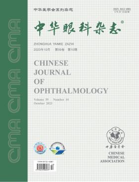
Volume 59 · Issue 10 · 2023
Multilingual International Publishing Platform for Humanities and Social Sciences111
This issue focuses on the research progress of corneal and ocular surface diseases. The responsible editorial committee responsible for the planning, composition and review of this issue is Professor Wang Liqiang.
In recent years, precision minimally invasive surgery has emerged as a significant developmental direction within the field of ophthalmology, bringing about revolutionary changes in the treatment of corneal diseases. Employing cutting-edge techniques and surgical methods, corneal surgeries have achieved greater precision and safety. In cases such as immune-mediated corneal diseases, it remains crucial to underscore the enhancement of vision through minimally invasive surgical approaches, thereby avoiding potential irreversible complications that surgery might induce. This article aims to assist readers in comprehending the profound impact of precision minimally invasive surgery on the treatment of corneal diseases, and calls upon medical professionals and researchers to continually explore and refine the realm of precision minimally invasive corneal surgery, fostering its further advancement.
Amniotic membrane (AM) can promote epithelialization, inhibit inflammation and be antimicrobial. It has been widely used in the clinical treatment of corneal epithelial and stromal defects. However, the misuse and abuse of AM might aggravate the primary disease and even lead to the loss of the best timing for other surgical interventions. Based on domestic and foreign clinical practice guidelines and medical evidence, the Cornea Group of Ophthalmology Branch of Chinese Medical Association has formed consensus opinions about the indications, surgical techniques and perioperative management of AM transplantation, so as to standardize the application of AM in the treatment of corneal and ocular surface diseases.
Abnormal visual experience in the first 3 years of life may lead to permanent visual impairment without early screening and timely intervention. It is of great clinical significance to evaluate the visual acuity of infants and preverbal children with suspected visual abnormality. However, the evaluation is difficult due to the inability of these children to express their complaints and the lack of unified standards. In order to promote the standardization of the visual acuity testing in infants and preverbal children, including the screening and follow-up examinations, a consensus has been reached by the Chinese Association for Pediatric Ophthalmology and Strabismus along with the Pediatric Ophthalmology and Strabismus Group of Chinese Ophthalmologist Association, based on the research results of evidence-based medicine.
To compare the efficacy and safety of a novel customized topography-guided transepithelial corneal collagen cross-linking (TG-CXL) procedure by sequential ultraviolet A irradiation in different diameters and conventional transepithelial corneal collagen cross-linking (TE-CXL) in adult patients with progressive keratoconus.
A prospective cohort study was conducted. Adult patients diagnosed with progressive keratoconus in the Affiliated Xiamen Eye Center of Xiamen University were continuously recruited and randomly assigned to receive the TG-CXL or TE-CXL procedure from March 2020 to March 2021. Patients in the TE-CXL group were irradiated in the central 9-mm zone of the cornea (total energy, 7.2 J/cm2; irradiance, 45 mW/cm2), while patients in the TG-CXL group were first irradiated with the protocol used in the TE-CXL group, and further irradiated in the central 6-mm zone (total energy, 3.6 J/cm2; irradiance, 9 mW/cm2). The subjective symptom of pain and corneal fluorescein sodium staining were scored within postoperative 3 days. Slit lamp examination, measurements of uncorrected visual acuity (UCVA) and best-corrected visual acuity (BCVA), corneal topography, anterior segment optical coherence tomography, in vivo corneal confocal microscopy, corneal endothelial cell count, and non-contact tonometry were performed before surgery and at 3, 6, and 12 months after surgery.
A total of 66 patients were enrolled (mean age, 23.0±3.3 years old), with 33 patients (33 eyes) in each group. No statistically significant differences were found in age, gender, and maximum keratometry (Kmax) between the two groups (P>0.05). On day 1 after surgery, the average pain score of the TG-CXL group (2.21±0.45) was significantly higher than that of the TE-CXL group (1.32±0.33) (P<0.05). The pain was rapidly alleviated in both groups on days 2 and 3. On days 1 and 2, the corneal fluorescein sodium staining scores in the TG-CXL group (4.15±0.83 and 2.21±0.60, respectively) were significantly higher than those in the TE-CXL group (1.76±0.56 and 0.85±0.51, respectively, P<0.001), while there was no significant difference between the two groups at day3 (P=0.184). The UCVA and BCVA of the TG-CXL group at 3, 6, and 12 months after surgery were significantly improved when compared with the baseline. At 3, 6, and 12 months, the BCVA (LogMAR) of the TG-CXL group (0.21±0.15, 0.22±0.16, and 0.22±0.16, respectively) were significantly improved when compared with those of the TE-CXL group(0.32±0.15, 0.34±0.15, and 0.36±0.16, respectively, P<0.01). However, there was no significant difference in UCVA between groups at any time point after surgery (P>0.05). The spherical and cylindrical power values of the TG-CXL group were improved when compared with the baseline (P<0.05). However, no significant difference in spherical power values was found between the two groups at any time point after surgery (P>0.05). Meanwhile, there were significant differences in cylindrical power values between the two groups at 6 and 12 months after surgery (P<0.05). The Kmax in the TG-CXL group was improved at all of the time points after surgery when compared with the baseline (P<0.001), while no significant difference in Kmax was found at any time point after surgery in the TE-CXL group when compared with the baseline (P>0.05). At 6 and 12 months after surgery, the Kmax values in the TG-CXL group were significantly lower than the TE-CXL group (P<0.05). No significant differences were found in flat keratomety, steep keratometry, the minimal thickness of the cornea, endothelial cell density, and intraocular pressure between the two groups at any time point after surgery (P>0.05). Within one month after surgery, optical coherence tomography revealed the increased density in the anterior stroma in both groups. In most patients in the TG-CXL group, a demarcation line was visible in the central and para-central corneal stroma, representing a clear and continuous, high-signal arc-shaped linear structure, which was deeper in the central cornea than the para-central cornea. In contrast, a demarcation line, fuzzy and focally discontinuous, was visible only in a few patients in the TE-CXL group, with an almost uniform depth in the central and the para-central cornea. Confocal microscopy demonstrated an apparent mesh-like cross-linked collagen structure in the superficial and intermediate corneal stroma at all time points after surgery in the TG-CXL group, with thickening stromal collagen fibers and an increased number of interconnections. In contrast, the mesh-like structure and number of interconnections in the superficial corneal stroma were significantly reduced at 12 months after surgery in the TE-CXL group, with no cross-linking structure in the intermediate corneal stroma at any time point after surgery. No serious complications such as corneal infection, sterile corneal ulcer, and persistent epithelial defect were observed in both groups during the follow-up of 12 months.
The TG-CXL procedure by sequential irradiation in two different diameters with ultraviolet A light was effective and safe in the management of progressive keratoconus in adults, achieving significant refractive improvement. This might be a good technical alternative for refractive corneal cross-linking surgery.
To evaluate the effectiveness and safety of 0.05% cyclosporine A and 0.1% tacrolimus eye drops in treating severe dry eye associated with chronic graft-versus-host disease (cGVHD).
This non-randomized concurrent control trial enrolled 83 eyes from 83 patients with cGVHD-associated severe dry eye. The treatment had two phases. During the initial shock treatment period (0-3 months), 44 patients received 0.05% cyclosporine A eye drops (4 times/day; group A) and 39 patients received 0.1% tacrolimus eye drops (twice/day; group B) alongside basic treatment. In the maintenance treatment period (3-6 months), both groups used 0.05% cyclosporine A eye drops (twice/day) and sodium hyaluronate. Examinations were conducted at 1, 3, and 6 months after treatment initiation, assessing the Ocular Surface Disease Index (OSDI), corneal fluorescein staining (CFS) score, and fluorescein tear break-up time (BUT) for efficacy. visual acuity and intraocular pressure (IOP) were evaluated for safety, and patients′ post-medication irritation symptoms were recorded.
The study included 52 males and 31 females, aged (28.57±15.67) years. After 1 month of treatment, the CFS score in group A significantly decreased from 10.0 (6.0, 14.0) to 5.0 (3.0, 8.5) (P<0.001). in group B, the CFS score also significantly decreased from 10.0 (6.0, 15.0) to 6.0 (2.0, 10.0), and the BUT increased from 2.0 (1.0, 2.0) s to 2.0 (1.8, 3.3) s (P<0.001). No significant OSDI decrease was observed in either group. No significant differences were found in OSDI, CFS score, and BUT between the two groups. After 3 months, group A showed significant improvement in OSDI, CFS score, and BUT (P<0.05), while group B only demonstrated significant CFS score decrease (P<0.05). OSDI was significantly lower in group A than group B (P<0.05). No significant differences were noted in CFS score and BUT between groups. After 6 months, OSDI, CFS score, and BUT were 18.9 (9.3, 34.2), 7.0 (3.0, 8.5), and 2.0 (1.0, 3.0) s in group A, and 10.9 (3.6, 35.4), 5.5 (2.8, 10.0), and 2.0 (1.0, 10.0) s in group B. In both groups, CFS scores significantly decreased and BUT increased (P<0.05). Visual acuity improved significantly in group A at 1, 3, and 6 months (P<0.05), while no significant changes were seen in group B. Irritation symptoms were transient and self-resolving in both groups.
Both 0.05% cyclosporine A and 0.1% tacrolimus eye drops, when combined with local glucocorticoids, exhibited significant anti-inflammatory effects, effectively and safely treating severe dry eye in cGVHD patients. Although the onset of 0.05% cyclosporine A was slower than 0.1% tacrolimus, it offered more stable long-term effects and better symptom improvement.
To explore the factors influencing the quality of donor corneal endothelium.
A retrospective case series study was conducted. Data from 568 donor corneas obtained from the Shandong Eye Bank between July 1, 2020, and June 30, 2021, were collected for analysis. The corneal endothelium of the donor corneas was observed using corneal endothelial microscopy to assess corneal endothelial cell density (ECD), coefficient of variation, and hexagonal cell ratio (HEX). Relevant factors of corneal donors were collected, including gender, age, cause of death, season of death, time from death to corneal retrieval, and methods of corpse preservation, to investigate their impact on the quality of donor corneal endothelium. The age factor was divided into five age groups: 0-20 years, 21-40 years, 41-60 years, 61-80 years, and >80 years. The time of corneal retrieval was divided into three periods based on the time elapsed since the donor's death: <6 hours, 6-12 hours, and >12 hours. The relationship between these factors and corneal endothelial conditions was analyzed.
The 568 donor corneas were obtained from 288 donors, including 225 males (78.13%) and 63 females (21.87%). The mean age was 51.77±18.48 years. The causes of death among donors were as follows: cardiovascular diseases 54.58% (275 individuals), cancer 17.96% (74 individuals), organ failure 14.26% (49 individuals), and accidents 13.20% (64 individuals). The mean time of corneal retrieval after donor death was 140 (76, 400) minutes (ranging from 30 minutes to 45 hours). Among the 145 corneas (25.53%) that had their initial corneal endothelial microscopy examination, the images were not clear, and after thorough rewarming, 106 corneas (18.7%) still had unclear images and could not be analyzed. Among the 462 corneas (81.3%) with clear images, the ECD was (2 602.23±318.40) cells/mm², the coefficient of variation was 36.61%±4.81%, and the HEX was 52.73%±7.15%. The ECD of corneas from older donors was lower compared to younger donors, and the differences between age groups were statistically significant (P<0.001). Corneas from donors who died due to accidents had a higher ECD [(2 829.88±313.90) cells/mm²] compared to those who died from cancer, cardiovascular diseases, and organ failure, and the differences were statistically significant (P<0.001). The ECD was highest when corneas were retrieved within 6 hours after death, and the difference was statistically significant (P<0.001). Older donors had higher coefficients of variation but lower HEX values (both P<0.05). Corneas retrieved after a longer time from death had higher coefficients of variation, and the difference was statistically significant (P<0.05), but there was no statistically significant difference in HEX (P>0.05). Organ failure, cryopreservation, and corneal retrieval time >12 hours were risk factors for unclear corneal endothelial imaging (all P<0.001). Among the 136 corneal endothelial images (23.94%), circular, oval, or band-shaped dark areas were observed, and corneas with dark areas had lower ECD (P<0.05). The longer the time elapsed from death to corneal retrieval, the more dark areas were observed (P<0.001). The presence of dark areas did not affect the coefficient of variation and HEX (P>0.05).
Advanced donor age, death due to chronic diseases, longer time elapsed from death to corneal retrieval, and cryopreservation of the body lead to a decrease in the quality of donor corneal endothelium.
To investigate the long-term outcomes of corneal grafts after penetrating keratoplasty(PK) for congenital corneal opacity(CCO) in children aged 0 to 5 years and the related influencing factors.
It was a retrospective series case study. Data of 39 children (55 eyes) who underwent PK surgery due to CCO in the keratology Department of Beijing Tongren Hospital from April 2014 to April 2018 and were followed up for more than 30 months were collected. Among them, there were 17 males (43.6%) and 22 females (56.4%). The age at operation was (16.2±13.3) months, and the follow-up time was (46.4±13.8) months. Clinical data such as basic information, preoperative diagnosis, operation age, operation method and postoperative complications were recorded. The corneal graft transparency was analyzed according to preoperative diagnosis, corneal neovascularization area, age at surgery, monocular or binocular surgery interval, primary surgery type and further surgery, and postoperative complications were observed.
At 12 months, 24 months and the last follow-up after PK, 78.2% (43/55), 70.9% (39/55) and 58.2% (32/55) of the affected eyes had clear corneal grafts, respectively.There was no statistical significance between Peters anomaly and sclerocornea (P>0.05), while the extent of neovascularization in the limbus had a significant effect on corneal graft transparency, and graft opacity was more likely to occur in patients with vessel area exceeding 2 quadrants (P<0.05).The highest corneal graft transparency was found in children aged 1 to 3 years 80.8%(21/26) (P<0.05), followed by children younger than 6 months (7/15).The translucency rate of the corneal graft was higher in patients undergoing unilateral surgery than in those undergoing bilateral surgery (P<0.05).Translucency of corneal graft was higher in children with simple surgery than with combined surgery (P<0.05), however, cataract surgery after PK had no significant effect on corneal graft transparency (P>0.05).The postoperative complications mainly included immune rejection in 19 eyes (34.5%), complicated cataract in 13 eyes (23.6%), glaucoma in 7 eyes (13.2%), persistent corneal epithelial defect in 7 eyes (13.2%).
After PK in children with CCO, the transparent rate of corneal grafts decreases gradually with time, but the long-term translucency of corneal grafts can still be obtained. The range of corneal neovascularization, age at the time of surgery, whether the surgery was binocular and whether the surgery was combined had an effect on the transparency of corneal graft.
To investigate the efficacy of Femtosecond laser-assisted stromal lenticule addition keratoplasty (SLAK) combined with corneal collagen cross-linking (CXL) in the treatment of middle and advanced Keratoconus.
It was a retrospective case series study. Data of 23 cases (24 eyes) of keratoconus treated with femtosecond laser-assisted SLAK combined with CXL in Laser Vision Centre of Xi′an No.1 Hospital from September 2020 to June 2022 were collected, including 16 males and 7 females, aged (23.69±5.18) years. The thickness, diopter number and diameter of the donor corneal stromal lens were assessed. uncorrected visual acuity (UCVA), best corrected visual acuity (BCVA), and diopter were recorded before and 1, 3, and 6 months after surgery. Sirius 3D fault corneal topography instrument to measure flat simulated keratometry (Kf), steep simulated keratometry (Ks) and the difference between them (ΔK), as well as central corneal thickness (CCT) and corneal high-order aberration.
Six months after surgery, CCT (454.83±50.01) μm were significantly higher than before (384.92±35.45) μm (P<0.05). Six months after surgery, UCVA (1.41±0.32) was significantly lower than before (1.11±0.33)(P<0.05). Six months after surgery, spherical diopter [(-15.73±7.89) D], Kf [(56.82±4.76) D] and Ks [(61.00±4.70) D] were significantly higher than before [(-12.08±5.99) D, (53.55±4.95) D, (58.65±5.10) D] (P<0.05). There was no significant difference in BCVA, column mirror degree and higher order aberrations before and 6 months after surgery(P>0.05). No corneal stromal lens folds, melting and displacement were observed in all eyes during the follow-up period, and no corneal opacity or immune rejection was observed.
femtosecond laser-assisted SLAK combined with CXL can significantly increase the corneal thickness of keratoconus and has good effectiveness. In addition, six months of postoperative follow-up of patients showed no significant changes in BCVA and high-order aberrations in the 6 mm central diameter of the cornea, and no postoperative adverse reaction were found in all eyes, indicating that the operation has certain safety.
To investigate the clinical presentation pattern of acute primary angle-closure glaucoma (PACG) during the 2019 novel coronavirus (2019-nCoV) pandemic over the past three years, and its relationship with 2019-nCoV infections of Omicron variants in Guangdong province.
Ecological study.Patients who were newly diagnosed with acute PACG from February 2020 to January 2023 at the Zhongshan Ophthalmic Center of Sun Yat-sen University were included in the study, and their basic information was collected. Patients were divided into the 2020 group (diagnosed between February 1st, 2020 and January 31st 2021), the 2021 group (diagnosed between February 1st, 2021 and January 31st 2022), and the 2022 group (diagnosed between February 1st, 2022 and January 31st 2023). The clinical presentation pattern of newly diagnosed acute PACG was observed and compared between groups. The daily number of newly diagnosed 2019-nCoV infections in Guangdong province was obtained from the Chinese Center for Disease Control and Prevention. The correlation between the daily number of newly diagnosed acute PACG and that of newly diagnosed 2019-nCoV infections during the epidemic period of Omicron variants between December 2022 and January 2023 was assessed.
The study included 1 048 patients with newly diagnosed acute PACG, with 235 for the 2020 group, 274 for the 2021 group, and 539 for the 2022 group. Our results showed that the average weekly number of newly diagnosed acute PACG patients in 2022 [8 (5, 11)] was significantly larger than that in 2020 (4.52±1.95, P<0.05) and 2021 (5.27±2.76, P<0.05). The average weekly number increased to 22.11±20.84 between December 2022 and January 2023. The total number of newly diagnosed acute PACG patients during this period was 199, which was 36.9% (199/539) of the total number of the same year and was 6.63 and 6.42 times as many as that in the same period (December and January) of 2020 and 2021. The proportion of patients with bilateral eye involvement during this period in 2022 was significantly higher than that in 2020 and 2021 (P<0.05). Further analysis found that 88.6% (109/123) of cases had a history of 2019-nCoV infection 2 (0, 3) days before the onset of acute PACG symptoms in average. The estimated daily number of acute PACG onset increased rapidly, peaked on December 23th, 2022, and then dropped gradually. This trend was similar to that of the daily number of new 2019-nCoV infections in Guangdong province. Changes of the daily number of new 2019-nCoV infections in Guangdong province had a positive correlation with the estimated daily number of acute PACG onset (r=0.84, P<0.001).
A dramatic increase in the clinical presentation of acute PACG was observed at Zhongshan Ophthalmic Center between December 2022 and January 2023, which was the epidemic period of Omicron variants. There is a correlation between the trend of the estimated daily number of acute PACG onset and that of new 2019-nCoV infections of Omicron variants in Guangdong province, but the exact reason remains to be further studied. (This article was published ahead of print on the official website of Chinese Journal of Ophthalmology on August 31, 2023)
A 60-year-old woman presented with a history of "pathological myopia combined with macular hole retinal detachment, " for which she underwent vitrectomy, internal limiting membrane tamponade, and silicone oil filling surgery one year ago. Seven months ago, the silicone oil was removed. She now returned to seek medical attention as her visual acuity has declined by 10 diopters. Examination revealed a recurrence of macular hole retinal detachment. The patient was treated with combined amniotic membrane tamponade and silicone oil filling. During the surgery, the frozen amniotic membrane was cut into 2 mm × 2 mm and placed under the macular hole retina after silicone oil filling. Within 6 months after the surgery, her retina repositioned well, the macular hole closed, and her visual function improved from hand motion to 0.05. No severe complications were observed. Therefore, this surgical approach provides a new perspective for treating recurrent pathological myopia combined with macular hole retinal detachment.
The patient was a 38-year-old female. Because a foreign body was splashed into his left eye for half a year, the anterior chamber disappeared for 1 d after the foreign body was taken from the outside hospital, and he went to Beijing Tongren Eye Center. Eye examination: visual acuity was 1.0 for the right eye and 0.6 for the left eye; IOP was 14 mmHg (1 mmHg =0.133 kPa) in the right eye and 6 mmHg in the left eye; No obvious abnormalities were observed in the anterior segment of the right eye; The conjunctiva of the left eyeball is mildly congested, the central cornea is flaky and opaque, and a round full-thickness defect with a diameter of about 1 mm can be seen at the temporal pupil margin corresponding to the cornea, with yellow deposits embedded around it. The anterior chamber is extremely shallow and the lens is transparent (Quintessential Picture 1); No obvious abnormalities were found in the fundus of both eyes. Clinical diagnosis: Penetrating corneal injury of left eye; Postoperative corneal foreign body removal in the left eye.
Anterior segment optical coherence tomography (AS-OCT) has the characteristics of non-contact, noninvasive, high sensitivity, and repeatability, and offers high-resolution in vivo imaging of the structures of the anterior eye segment. It can be used in the diagnosis and assistance of conditions related to the anterior eye segment. This review provides an update on the research and clinical applications of AS-OCT in corneal refractive surgery, including preoperative keratoconus screening, intraoperative real-time visualization of corneal structures, postoperative corneal evaluation, and management of postoperative complications. We also explore the potential application of AS-OCT in combination with corneal biomechanical detection for corneal refractive surgery.
Acanthamoeba keratitis (AK) is an infectious keratitis with difficulty in early diagnosis, poor curative effect, high blindness rate and serious threat to the visual acuity of patients. Delayed diagnosis can lead to prolonged course of AK, loss of vision, increased probability of corneal transplantation, and some patients even need to remove eyeballs. Therefore, early diagnosis is related to the visual prognosis of patients. This article starts with the risk factors, early clinical manifestations, auxiliary examinations and results of AK, and puts forward the process and key points of early diagnosis and differential diagnosis of AK, in order to improve the clinical diagnosis level, realize the early diagnosis of AK, and thus save the visual acuity of AK patients to the greatest extent.
CURRENT ISSUE