Multilingual International Publishing Platform for Humanities and Social Sciences111
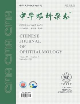
Volume 59 · Issue 09 · 2023
Multilingual International Publishing Platform for Humanities and Social Sciences111
This issue focuses on the research progress of glaucoma. The responsible editorial committee responsible for the planning, composition and review of this issue is Professor Zhang Chun.
Compared to traditional filtration surgery, minimally invasive glaucoma surgery (MIGS) offers several advantages, including smaller incisions, more effective intraocular pressure reduction, higher safety levels, fewer surgical complications, faster postoperative recovery, and reduced postoperative use of antiglaucoma drugs. MIGS has gained rapid popularity both domestically and internationally due to its easier operation, shorter learning curve, and reduced operation time. Considering the current clinical promotion of MIGS and the application of health economic evaluation in the glaucoma field, this article proposes the need for an emphasis on the health economic evaluation of MIGS. Conducting research in this area will provide valuable insights for related clinical diagnosis and treatment, medical pricing, and medical insurance payment, and help to further disseminate MIGS, so as to offer clinical benefits, which are aligned with national conditions and economic viability.
The rapid iteration of artificial intelligence (AI) technology has driven the development of intelligent ophthalmology, with enormous potential for application in the regional screening, precise diagnosis, and disease progression prediction of intractable blinding diseases, particularly glaucoma. Aiming to encourage collaborative discussions among ophthalmologists, this article elucidates the application potential and direction of AI in the prevention and treatment of glaucoma from the aspects of enhancing glaucoma screening and diagnosis levels through AI, promoting initiatives for the application of AI, and exploring the prospects of AI.
Minimally invasive glaucoma surgery (MIGS) is widely used in China because of its effective reduction of intraocular pressure, small incisions, few surgical complications, and quick postoperative recovery. In order to regulate and promote the clinical application of MIGS, the Glaucoma Group of Ophthalmology Branch of Chinese Medical Association has formed consensus opinions on the clinical application and perioperative management of MIGS with subconjunctival implants on the basis of domestic and foreign research results and clinical practice, providing reference and guidance for clinical work.
To compare the efficacy and safety of trabeculotome tunnelling trabeculoplasty and gonioscopy-assisted transluminal trabeculotomy (GATT) in the treatment of open-angle glaucoma.
A prospective randomized controlled study. The patients with open-angle glaucoma diagnosed in the ophthalmology center of Beijing Tongren Hospital affiliated to Capital Medical University from January to July 2022 were collected and divided into GATT group (undergoing GATT) and 3T group (undergoing 3T operation) using a random number table. Intraocular pressure (IOP) was recorded for both groups at 1 day, 1 week, 1 month, and 3 months after the operation, and the types and quantities of anti-glaucoma drugs used, postoperative complications, and surgical success rate were compared. Normal distribution measurement data were analyzed using independent sample t-tests, non-normal distribution measurement data were analyzed using non-parametric tests, and counting data were analyzed using chi-square tests.
This study included 35 patients (43 eyes), consisting of 27 males and 8 females, with an average age of (43.0±14.3) years. There were 21 patients (23 eyes) in the GATT group and 19 patients (20 eyes) in the 3T group. The maximum IOP without anti-glaucoma drugs before surgery, the highest IOP with the maximum number of anti-glaucoma drugs, and the IOP at 3 months after surgery in the GATT group were (33.5±9.1), (22.2±6.1), and (16.0±3.1) mmHg (1 mmHg=0.133 kPa), respectively. The corresponding values for the 3T group were (35.2±7.8), (21.5±6.8), and (16.1±2.0) mmHg. After surgery, the IOP in both groups was lower than before surgery, with a statistically significant difference (P<0.05) and no significant difference between the two groups (P>0.05). In the 3 months following surgery, 13 eyes in the GATT group and 11 eyes in the 3T group received more than two types of anti-glaucoma drugs, with no significant difference between the two groups (P>0.05). Three months after surgery, the complete and conditional success rates of the GATT group were 14/18 and 16/18, respectively, and those of the 3T group were 12/15 and 13/15, respectively, with no significant difference between the two groups (P>0.05). The incidence of hyphema, ciliary detachment, and shallow anterior chamber 1 day after surgery was 91%(21/23), 35%(8/23), and 30%(7/23), respectively, in the GATT group and 55%(11/20), 5%(1/20), and 0 in the 3T group, with a statistically significant difference between the two groups (P<0.05).
3T and GATT have similar success rates in the treatment of open-angle glaucoma. However, compared with GATT, 3T has fewer complications and is considered to be safer. (This article was published ahead of print on the Online-First Publishing Platform for Excellent Scientific Researches of Chinese Medical Association Publishing House on February 28, 2023)
To analyze the disease composition and primary surgical procedures in pediatric inpatients with secondary glaucoma.
A retrospective case series study was conducted. Clinical data of children aged≤16 years with secondary glaucoma who were admitted to the Zhongshan Ophthalmic Center, Sun Yat-sen University, between January 1, 2017, and December 31, 2021, were included. The patients were classified according to the Childhood Glaucoma Research Network (CGRN) classification system, and their diagnoses, underlying factors, gender, age of onset, affected eye(s), age and type of initial surgery, and ophthalmic examination data were analyzed. Statistical analysis was performed using Kruskal-Wallis rank sum test and χ2 test.
A total of 540 patients (744 eyes) were included in this study, comprising 319 males (59.1%) and 221 females (40.9%). Unilateral disease was observed in 336 cases (62.2%), while bilateral involvement was present in 204 cases (37.8%). The age of onset was 4.0 (0.0, 9.0) years, and the median age of the first anti-glaucoma surgery was 5.0 (0.7, 10.0) years. Among them, there were 195 cases (36.1%) of secondary glaucoma associated with non-acquired ocular anomalies (SCG-O), with a median age of onset of 0.0 (0.0, 4.0) years, and 97 of these cases (49.7%) were male. secondary glaucoma associated with non-acquired systemic disease or syndrome (SCG-S) were observed in 68 cases (12.6%), with a median age of glaucoma onset of 0.1 (0.0, 4.0) years, and 47 of these cases (69.1%) were male. Secondary glaucoma associated with acquired conditions (SCG-A) accounted for 192 cases (35.6%), with a median age of onset of 9.0 (5.0, 13.0) years, and 125 of these cases (65.1%) were male. There were 85 cases (15.7%) of secondary glaucoma following cataract surgery (SCG-C), with a median age of onset of 3.0 (0.8, 7.0) years, and 50 of these cases (58.8%) were male. Male patients were predominant in SCG-S and SCG-A, with 47 cases (69.1%) and 125 cases (65.1%), respectively (χ2=9.94, 17.52; P=0.002,<0.001). Except for SCG-O, all other types of pediatric secondary glaucoma predominantly affected only one eye: SCG-S in 52 cases (76.5%), SCG-A in 128 cases (66.7%), and SCG-C in 54 cases (63.5%) (χ2=19.06, 21.33, 6.22; all P<0.05). The highest proportion of SCG-O was attributed to congenital ectropion uveae (46 cases, 23.6%). Sturge-Weber syndrome was the most common SCG-S (45 cases, 66.3%), while SCG-A mostly resulted from trauma (59 cases, 30.8%) and corticosteroid use (56 cases, 29.2%). Trabeculectomy (211 eyes, 30.8%) and glaucoma drainage device implantation (197 eyes, 28.7%) were the most frequently performed primary surgical procedures.
SCG-O and SCG-A were found to be common types of pediatric secondary glaucoma. The age of onset and the choice of primary anti-glaucoma surgical procedures varied among different types of pediatric secondary glaucoma. However, overall, trabeculectomy and glaucoma drainage device implantation were the primary surgical procedures predominantly employed.
To evaluate the medium-term efficacy and safety of Ex-PRESS shunt implantation in the treatment of secondary glaucoma associated with Sturge-Weber Syndrome (SWS).
This was a retrospective case series study. Medical records of patients diagnosed with secondary glaucoma due to SWS who underwent Ex-PRESS shunt implantation at Shanghai Ninth People′s Hospital, Shanghai Jiao Tong University School of Medicine, between January 2013 and February 2020 were collected. Only the right eye were included in the analysis when both eyes were affected. Follow-up visits were scheduled at postoperative week 1, 3 months, 6 months, 1 year, 2 years, and 3 years. Clinical data including intraocular pressure (IOP), vertical cup-to-disc (C/D) ratio, corneal horizontal diameter, visual field, anterior chamber condition, anti-glaucoma medication usage, and additional surgeries were evaluated and analyzed before and after the procedure. Surgical success rates and procedure-related complications at each follow-up time point were assessed. Statistical analyses were performed using t-test, Mann-Whitney U test, and χ2 test.
A total of 21 patients (21 eyes) were included in the study, comprising 10 females and 11 males. Among them, 15 eyes were on the right side, and 6 eyes were on the left side. The patients′ ages ranged from 3 to 51 years, with a median age of 8.1 (6.3, 11.9) years. The follow-up period ranged from 3 to 53 months, with a median of 11 (6, 24) months. Preoperatively, the IOP in the 21 operated eyes was (32.9±9.1) mmHg (1 mmHg=0.133 kPa), significantly higher than the IOP in the 19 healthy eyes, which was (17.1±4.3) mmHg (t=5.80, P<0.001). The C/D ratio in the operated eyes was (0.75±0.13), also significantly higher than that in the healthy eyes, which was (0.32±0.10) (t=11.22, P<0.001). At the 1-year, 2-year, and 3-year follow-up, 8 eyes out of 16 operated eyes, 6 eyes out of 9 operated eyes, and 7 eyes out of 10 operated eyes achieved overall surgical success (complete success+conditional success), respectively. The number of eyes with complete success at the three follow-up time points was 3, 2, and 4, respectively. The IOP in the operated eyes was significantly reduced at all follow-up time points compared to preoperative values (all P<0.05), while there was no statistically significant difference in C/D ratio before and after surgery (all P>0.05). Two operated eyes experienced retinal detachment or choroidal leakage postoperatively, both of which recovered after conservative treatment. One eye developed postoperative degree Ⅰ shallow anterior chamber, and it resolved spontaneously on the third day after surgery. No serious surgical complications, such as bleb-related complications, malignant glaucoma, expulsive choroidal hemorrhage, or endophthalmitis, were observed postoperatively.
Ex-PRESS shunt implantation for the treatment of secondary glaucoma in SWS demonstrated a relatively high level of safety. The medium-term IOP in the operated eyes significantly decreased compared to preoperative values. However, the majority of operated eyes did not achieve complete surgical success.
To evaluate the clinical application value of intraoperative optical coherence tomography (iOCT) in deep anterior lamellar keratoplasty (DALK) using the big-bubble technique to bare Descemet′s membrane.
Retrospective case series. Clinical data of 92 patients (92 eyes) with monocular stromal corneal diseases who underwent big-bubble DALK in the Eye Hospital of Shandong First Medical University from January 2020 to August 2021 were collected. There were 53 males and 39 females. The average age was (53.2±16.0) years old. All patients underwent iOCT scanning to determine the location and depth of the injection needle after initial removal of the corneal lesion, to observe the integrity of the recipient bed, Descemet′s membrane, after complete lesion removal, and to observe the adhesion between the corneal graft and the recipient bed and check folds on the recipient bed after suturing of the corneal graft. The intraoperative perforation of Descemet′s membrane, postoperative thickness of the cornea and the recipient bed, visual acuity, and corneal astigmatism were recorded.
By iOCT, the thickness of the recipient bed was found to be about 1/2 of the corneal thickness and relatively uniform in all directions in 62 eyes (67.4%), so the sterile air was injected from the center of the recipient bed to separate it from the stromal layer. In 30 eyes (32.6%) with an uneven thickness of the recipient bed, the sterile air was injected from the paracentral area of the recipient bed. Under the guidance of iOCT scanning, 89 eyes (96.7%) did not experience any perforation of Descemet′s membrane during surgery. The Descemet′s membrane folds in the central 5-mm area of the recipient bed was observed and flattened in 20 eyes with the assistance of iOCT scanning. The postoperative corneal thickness was (578.95±108.26) μm, and the recipient bed thickness was (36.06±23.11) μm. The best corrected visual acuity of all patients at 6 months after surgery was 0.57±0.25 logMAR, which was significantly better than that before surgery (1.61±1.27 logMAR; P<0.001). The average corneal astigmatism at 6 months after surgery was (2.72±2.44) diopters.
The application of iOCT scanning in DALK surgery assisted by the big-bubble method can provide safe guidance for surgeons to adopt correct surgical procedures, decrease the risk of Descemet′s membrane perforation, reduce the recipient bed folds, and facilitate corneal interlayer adhesion, thereby improving the visual prognosis.
To investigate the effects and mechanisms of long-term high-fat diet on synaptic plasticity in the visual cortex and hippocampus neurons of juvenile mice.
This was an experimental study. Twenty-four 4-week-old male C57BL/6J mice were randomly divided into two groups, using a randomized numerical table, with 12 mice in each group. The ND group was fed a normal diet, while the HFD group was fed a high-fat diet. After 12 weeks of feeding, mouse body weight, body fat percentage, glucose tolerance, and blood lipid levels were recorded. Six mice from each group were randomly selected using a randomized numerical table, and long-term potentiation (LTP) in the lateral geniculate nucleus (LGN)-primary visual cortex binocular zone (V1B area) and hippocampus CA3-CA1 were recorded in vitro. Field excitatory postsynaptic potentials (fEPSPs) were measured, and the normalized fEPSP slope was calculated to evaluate changes in cortical synaptic plasticity. Subsequently, brain tissue was collected for Golgi staining to observe the development of pyramidal neurons in layers Ⅱ-Ⅲ of the primary visual cortex and CA1 region of the hippocampus, and changes in dendritic spine morphology and quantity were compared. The remaining six mice from each group were euthanized, and brain tissue was collected for transmission electron microscopy to observe ultrastructural changes in the visual cortex V1B area and hippocampus CA1 region neurons. Independent samples t-test was used for statistical analysis.
After 12 weeks of feeding, the body weight of mice in the HFD group was (29.17±1.63) g, significantly lower than the ND group which was (37.99±6.87) g (t=4.33, P<0.001). The body fat percentage in the HFD group was 1.09%±0.22%, which was higher than the ND group with 0.85%±0.09% (t=2.50, P=0.032). HFD mice showed a significant increase in blood glucose level 30 minutes after glucose injection, reaching (17.80±3.94) mmol/L, compared to the ND group with (23.10±1.48) mmol/L (t=3.07, P=0.013). At 60 minutes after glucose injection, the difference in blood glucose levels between the ND group [(13.58±2.39) mmol/L] and the HFD group [(23.70±3.56) mmol/L] was statistically significant (t=5.40, P<0.001). Subsequently, both groups showed a decline in blood glucose levels, and at 120 minutes after glucose injection, the blood glucose level in the ND group decreased to (8.50±1.05) mmol/L, while the HFD group remained at a higher level of (16.03±4.17) mmol/L, showing a statistically significant difference (t=3.91, P=0.004). The serum total cholesterol levels in the ND and HFD groups were (4.08±0.35) mmol/L and (10.80±0.90) mmol/L, respectively, with the HFD group higher than the ND group (t=15.23, P<0.001). However, there was no significant difference in triglyceride levels (P>0.05). The high-density lipoprotein cholesterol level in the ND group was (2.12±0.57) mmol/L, while in the HFD group, it was (1.28±0.15) mmol/L, with the HFD group lower than the ND group (t=3.15, P=0.014). Non-high-density lipoprotein cholesterol level in the HFD group was (11.06±1.46) mmol/L, significantly higher than the ND group with (2.28±0.43) mmol/L (t=12.88, P<0.001). In the hippocampal CA3-CA1 pathway, the fEPSP slope increased by 239.1%±88.8% of baseline in the ND group, while in the HFD group, it was only 147.6%±31.6% of baseline, indicating lower LTP compared to the ND group (t=7.20, P<0.001). For the LGN-V1 pathway, the fEPSP slope increased by 204.8%±67.0% of baseline in the ND group, while in the HFD group, it was 121.1%±15.7% of baseline, showing reduced LTP compared to the ND group (t=9.11, P<0.001). Regarding the visual cortex, in the V1B area of the ND group, the number of dendritic spines per 10 μm was (1.31±1.14), while in the HFD group, it was (0.77±0.43), demonstrating a significant decrease in dendritic spine density (t=3.45, P<0.001). The proportion of mature dendritic spines in the ND group was 69.98%, while non-mature dendritic spines accounted for 30.02%. In contrast, the HFD group had 45.76% mature dendritic spines and 54.24% non-mature dendritic spines. Regarding changes in hippocampal CA1 pyramidal neurons, the cell bodies and axons were not damaged, but HFD group neurons exhibited simplified dendritic structures with reduced branching. The number of dendritic spines per 10 μm was (10.25±3.84) in the HFD group and (25.22±8.21) in the ND group, indicating significantly lower dendritic spine density in the HFD group (t=12.42, P<0.001). The proportion of mature dendritic spines in the ND group was 70.88%, while non-mature dendritic spines accounted for 29.12%. In contrast, the HFD group had 47.37% mature dendritic spines and 52.63% non-mature dendritic spines. Moreover, the ultrastructure of neurons in the visual cortex V1B area and hippocampus CA1 region of HFD mice showed evident damage, with disrupted cell structures, swollen and vacuolated mitochondria, reduced or even disappeared mitochondrial cristae, and decreased synaptic quantity with damaged structure.
Long-term high-fat diet in juvenile mice leads to abnormal development and functional maturation of synapses in the visual cortex and hippocampal regions. Dendrites, as the foundation of synaptic structures, undergo abnormal development, which can cause alterations in synaptic plasticity of related neural circuits.
A 69-year-old female patient presented to the ophthalmology department with complaints of blurred vision in the left eye for more than 10 days. Her medical history revealed a history of right renal tumor and left pheochromocytoma, which were treated with surgical resection at an external institution. Ophthalmic examination revealed a temporal superior cup-shaped optic disc pit in the left eye, along with a macular hole approximately 1/5 the size of the optic disc diameter in the macular region. Additionally, peripheral retinal examination at the 6 o′clock and 11 o′clock positions showed vascular tumors, each approximately 1.5 times the size of the optic disc diameter. Based on the patient′s medical history, fundus findings, and auxiliary examination results, a diagnosis of macular hole in the left eye, optic disc pit in the left eye, and Von Hippel-Lindau (VHL) syndrome was established. Subsequently, the patient underwent left vitrectomy and macular hole repair surgery, leading to an improvement in visual acuity.
This case report presents a 5-year-old male child with a complaint of poor vision in the left eye for the past 2 years, who sought ophthalmic evaluation. There was no apparent systemic pigment loss, but multiple small, flat, and well-defined white lesions were observed in the retinal pigment epithelium of the left eye. Autofluorescence imaging of the fundus revealed widespread patchy hyperautofluorescence corresponding to the lesions. Fluorescein angiography demonstrated early and stable hyperfluorescence without leakage in these spots. Optical coherence tomography examination revealed thickening and material accumulation in the ellipsoid zone that corresponded to the lesions. Based on clinical findings, the diagnosis of congenital grouped albinotic spots was established.
A female patient, 87 years old, presented with an irregular swelling in the left medial canthus for 7 years. Due to the patient′s poor general condition, radical surgery was not considered appropriate. Surgical biopsy confirmed periocular basal cell carcinoma of the left medial canthus. The patient was administered oral HedgeHog inhibitor targeted therapy for 6 months, resulting in approximately 90% reduction in tumor size. The patient′s condition improved, and the tumor remained stable during the course of follow-up.
Surgeon introduction Wang Ningli, chief physician, professor. Director of Beijing Tongren Ophthalmology Center, Beijing Tongren Hospital affiliated to Capital Medical University, and Dean of College of Ophthalmology, Capital Medical University. Former chairman of Ophthalmology Branch of Chinese Medical Association and leader of glaucoma group, president of Ophthalmologist Branch of Chinese Medical Doctors Association, chairman of Public Health Ophthalmology Branch of Chinese Preventive Medicine Association, and leader of National Blindness Prevention Technical Guidance Group; Honorary Editor-in-Chief of the Editorial Board of Chinese Journal of Ophthalmology; Fellow of the International Academy of Ophthalmology, President of the Asia-Pacific Ophthalmology Society, Member of the Board of Directors of the World Glaucoma Association, and Member of the Board of Directors of the Asia-Pacific Glaucoma Association.
The patient was a 40-year-old male. Because of the discovery of conjunctival cyst in his left eye for half a year, he went to the Department of Ophthalmology, Peking University People's Hospital. Trabeculectomy of left eye was performed in external hospital 5 years ago for open angle glaucoma. Eye examination: corrected visual acuity was 0.6 for the right eye and 0.1 for the left eye; IOP was 15 mmHg (1 mmHg =0.133 kPa) in the right eye and 16 mmHg in the left eye; The left eye has a white cystic bulbous conjunctiva at the posterior scleral margin of the superior temporal angle, with a size of about 7 mm ×8 mm, and clusters of blood vessels are seen on the surface (essence picture 1). The root is connected to the filter vesicle, and it is soft in texture and has good mobility. The cornea of both eyes was transparent, the anterior chamber depth was normal, and the lens was mildly opaque; The optic disc boundary of both eyes is clear, the ratio of optic cup to optic disc diameter is about 0.5, and the retina is in place. The anterior segment coherence light tomography examination is shown in Picture Essence 2. Clinical diagnosis: Prolapse of conjunctival filtering vesicles after surgery for glaucoma filtering glaucoma in the left eye.
Selective Laser Trabeculoplasty (SLT) is an effective laser treatment modality for controlling intraocular pressure (IOP) in open-angle glaucoma (OAG). Compared to other laser techniques targeting the trabecular meshwork, SLT has demonstrated favorable efficacy and safety. This article aims to summarize the application and advancements of SLT in Chinese OAG patients, focusing on its effectiveness in reducing IOP and postoperative adverse reactions that may influence OAG. By examining SLT from these perspectives, we aim to provide insights for the standardized promotion of SLT and enhancement of clinical glaucoma management in China.
Myopia and glaucoma are two common ocular diseases that pose significant health and economic burdens to patients, their families, and society. In recent years, the relationship between these two conditions has become a prominent research focus. This article provides a systematic review of the current research progress on the association between myopia and glaucoma, with a primary emphasis on discussing the two major viewpoints: myopia as a risk factor for glaucoma development and the multifaceted impact of myopia on glaucoma progression. Possible mechanisms and potential clinical biomarkers are outlined, and the academic community′s complementary insights and doubts regarding these viewpoints are summarized. Additionally, unresolved issues in existing studies are identified to pave the way for future scientific explorations, aiming to further elucidate the relationship between myopia and glaucoma and optimize the clinical diagnosis and management of glaucoma.
Monocular elevation deficiency (MED) is a functional defect in upward eye movement caused by paralysis, restriction, or a combination of factors. The affected eye experiences limited movement in the upward, nasal, and temporal directions, often accompanied by downward deviation and ptosis. Previously known as double elevator palsy (DEP), MED can result from muscle paralysis responsible for upward movement, restriction of the inferior rectus muscle, or lesions in the supranuclear pathway. The Knapp procedure was once the most commonly used surgical approach for MED. However, in cases involving restriction of the inferior rectus muscle, a staged approach combining inferior rectus muscle weakening and subsequent Knapp procedure is necessary to avoid anterior segment ischemia. This has led to the development of alternative surgical techniques, such as single-rectus muscle transposition, partial tendon transposition, and muscle belly transposition. Differentiating MED subtypes based on the underlying paralysis or restriction etiology is crucial for determining the appropriate surgical plan. This review article summarizes the clinical features, classification, and available surgical options for MED, aiming to provide valuable insights for clinical research and treatment.
There are more than 20 causes that can cause angle closure in young people under 40 years old, of which primary angle closure glaucoma accounts for only about 1/3. Due to the different pathogenesis of various etiologies, the treatment and prognosis are far from each other, so it is necessary to conduct comprehensive clinical examination, careful screening and diagnosis, and careful choice of treatment plan. This paper introduces the etiology and diagnosis and treatment points of angle closure in young people in five categories, in order to provide reference for clinical work.
CURRENT ISSUE