Multilingual International Publishing Platform for Humanities and Social Sciences111
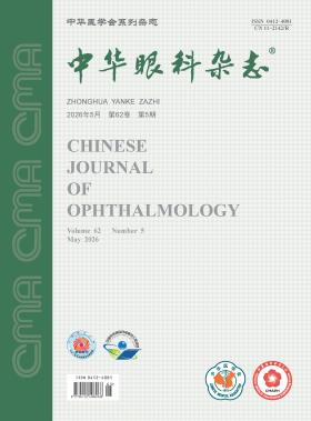
Volume 62 · Issue 05 · 2026
Multilingual International Publishing Platform for Humanities and Social Sciences111
This issue focuses on the research progress of strabismus and pediatric ophthalmology diagnosis and treatment. The responsible editorial committee responsible for the planning, composition and review of this issue is Professor Zhang Wei.
Children′s visual health serves as a strategic cornerstone for national health and human capital, exerting a lifelong influence that profoundly shapes individual development and social well-being. At present, visual impairment among Chinese children is characterized by a high overall prevalence and a complex, multifactorial etiology. Major conditions include refractive errors, amblyopia, strabismus, congenital cataract, congenital glaucoma, retinopathy of prematurity, and cerebral visual impairment. A substantial proportion of these impairments are preventable or curable; however, the current prevention and control system remains constrained by limited public awareness, insufficient alignment of screening, diagnostic and therapeutic technologies, and a fragmented service system. In response, this paper focuses on delineating the core etiologies and multidimensional health hazards of childhood visual impairment, identifying key bottlenecks in existing prevention and treatment practices, and exploring theoretically grounded and practically integrated strategies to establish a tiered, collaborative, and efficient prevention and control pathway. This effort aims to consolidate progress in addressing childhood visual impairment in China, reduce avoidable visual impairment, and ultimately improve the overall level of children′s visual health nationwide.
With the rapid development of generative artificial intelligence (AI) technologies and large AI models, these technologies are expected to play an important role in multiple aspects of medical workflows in ophthalmology. However, their rapid and widespread adoption has also introduced multiple challenges regarding application specifications, ethical constraints, and legal compliance. To ensure the safe, effective, and compliant application of generative AI large models in ophthalmology, the Intelligent Ophthalmology Subcommittee (China) of Technical Committee on Smart Medical Digitalization of IEEE Technology and Engineering Management Society convened domestic experts in the field. Drawing upon relevant Chinese laws and regulations governing medical practice, data security, and privacy protection, and incorporating the latest research advances, the expert panel held multiple online and offline seminars nationwide. Using the Grading of Recommendations Assessment, Development and Evaluation (GRADE) system, Good Practice Statements, and the Delphi method, the panel developed a consensus framework addressing the ethical, technical, and legal issues associated with the application of generative AI large models in ophthalmology. Designed to be continuously updated as technology advances, this framework aims to provide evidence-based support for clinical practice.
To evaluate the efficacy and safety of low-dose compound tropicamide eye drops for pupillary dilation in premature infants undergoing the fundus examination.
A non-inferiority, randomized, controlled, crossover trial was conducted. Preterm infants hospitalized in the Department of Neonatology, the First Affiliated Hospital of Zhengzhou University scheduled for retinopathy of prematurity screening between October 2024 and June 2025 were enrolled. Participants were randomly assigned to group A or group B using simple randomization. Group A received standard-dose (90 μl) compound tropicamide eye drops in the first fundus examination, but low-dose (45 μl) drops in the second examination, whereas group B received the two doses in the reverse order. The primary efficacy outcomes were the pupil diameter measured 60 to 75 minutes after dilation and the difficulty of fundus examination as assessed by the ophthalmologist (classified as easy, difficult, or impossible). The difference in pupil diameter between the two groups and its 95% confidence interval (CI) were calculated and compared with the non-inferiority margin to determine non-inferiority. The safety was assessed based on changes in vital signs and adverse events. A linear mixed model and generalized estimating equations were used to compare differences between groups.
A total of 72 infants were enrolled. Group A comprised 34 infants, of whom one was lost to follow-up during the second examination; group B comprised 38 infants, of whom three were lost to follow-up during the second examination. In the two groups, the gestational ages at birth were (30.85±2.38) and (31.81±2.61) weeks, the birth weights were (1 262.06±422.56) and (1 357.63±389.31) g, and male infants accounted for 35.29% (12 cases) and 50.00% (19 cases), respectively. No statistically significant differences were observed in demographic characteristics or clinical complications between the two groups (all P>0.05). At 60 to 75 minutes after dilation, the difference in pupil diameter between the low-dose and standard-dose regimens was 0.24 mm (95% CI: 0.18 to 0.29), with the upper limit of the 95% CI falling below the pre-specified non-inferiority margin. The rate of examination difficulty was 3% (95% CI: 1% to 11%) after standard-dose administration and 7% (95% CI: 3% to 16%) after low-dose administration, with a rate difference of 4% (95% CI: -1% to 9%, P=0.087; after Bonferroni correction, P=0.435). The lower limit of the 95% CI was above -5%. After low-dose administration, the oxygen saturation by pulse oximetry was 0.09% lower (95% CI: -0.32 to 0.15, P=0.470; after Bonferroni correction, P=1.000), and the heart rate was 0.34 beats/min lower (95% CI: -1.11 to 0.43, P=0.381; after Bonferroni correction, P=1.000), compared with standard-dose administration, with no statistically significant differences. The systolic blood pressure was 0.45 mmHg (1 mmHg=0.133kPa)lower (95% CI: -0.86 to -0.05, P=0.028; after Bonferroni correction, P=0.140), and the diastolic blood pressure was 0.65 mmHg lower (95% CI: -1.05 to -0.25, P=0.001; after Bonferroni correction, P=0.005) after low-dose administration compared with standard-dose administration, with only the difference in diastolic blood pressure reaching statistical significance. Adverse events occurred 11 times after standard-dose administration and 3 times after low-dose administration.
For fundus screening in preterm infants, low-dose (45 μl) compound tropicamide eye drops are non-inferior to the standard dose (90 μl) in terms of mydriatic efficacy, while offering advantages in safety profile.
To investigate the genetic characteristics of copy number variations (CNVs) in families with concomitant exotropia.
A family-based molecular genetic study. The study included 25 unrelated concomitant exotropia families who visited Tianjin Eye Hospital between August 2019 and August 2021. Patients and healthy individuals from these families were included as subjects. Clinical data were collected through detailed medical history interviews, family history documentation, and comprehensive ophthalmic and systemic examinations to confirm the diagnosis of probands and affected family members. Peripheral blood samples were obtained for genomic DNA extraction, followed by single nucleotide polymorphism (SNP) genotyping using single nucleotide polymorphism array (SNP array). CNV detection was performed using the PennCNV tool. Gene enrichment analysis and protein-protein interaction (PPI) network analysis were conducted to explore the biological functions of candidate genes and their potential pathogenic mechanisms. The Benjamini-Hochberg method was used for multiple comparison correction, and node permutation randomization test was used for significance assessment of network analysis.
A total of 74 subjects from 25 families with concomitant exotropia were enrolled in the study, including 55 patients with concomitant exotropia. Among all subjects, 33 were male (44.6%) and 41 were female (55.4%). Among the 55 patients with concomitant exotropia, 27 were male (49.1%) and 28 were female (50.9%). The age at onset was 6.0 (3.3, 10.0) years. Using the prism and alternate cover test, the deviation angle was -30Δ (-39Δ, -25Δ) at distance (6 m) and -35Δ (-40Δ, -30Δ) at near (33 cm). Based on the SNP genotyping technique, this analysis identified seven novel concomitant exotropia-associated CNVs, located at chromosomal regions 2p25.1, 20q13.33, 15q22.2, 7q34, 13q14.2, 8p21.1, and 3q25.33. Analysis of familial distribution showed that the CNV at 2p25.1 had the highest detection rate (28%), being present in seven families. Co-segregation analysis revealed that CNVs at 15q22.2, 7q34, and 13q14.2 exhibited complete co-segregation with the disease phenotype (100% penetrance), whereas regions at 2p25.1, 20q13.33, 8p21.1, and 3q25.33 showed incomplete penetrance of 73.3%, 92.3%, 83.3%, and 80%, respectively. These seven CNVs covered the exonic regions of nine protein-coding genes, including ROCK2, SYCP2, VPS13C, RB1, BRAF, CCDC25, ESCO2, PBK and IFT80. Gene enrichment analysis revealed that these genes were enriched in pathways associated with muscular movement regulation (q<0.001). Gene intersection analysis identified two genes (ESCO2 and BRAF) that overlap with genes related to oculomotor neural development, and four genes (ESCO2, BRAF, RB1 and IFT80) that overlap with genes related to extraocular muscle development. PPI analysis revealed that ROCK2, BRAF, RB1, PBK, and ESCO2 proteins formed a significant functional cluster with a higher number of edges than random expectation(μ=0.09±0.42,P=0.002,Z=9.31).
This study reports seven CNVs associated with concomitant exotropia. These CNVs exhibit genetic characteristics of scattered chromosomal distribution, variable penetrance and functional clustering of target genes.
To investigate the consistency of different optical coherence tomography (OCT) devices in measuring key retinal parameters.
This study consisted of a cross-sectional study and an experimental study. A total of five OCT devices of different brands (Optovue OCT, Topcon OCT, Intalight OCT, TowardPi OCT, and Zeiss Cirrus OCT) were included, and corresponding devices were selected according to different measurement objects and parameters. Retinal thickness and cup-to-disc ratio were measured in 8 eyes of 4 healthy volunteers recruited from the Department of Ophthalmology, Tianjin Medical University General Hospital, between May and August 2024. In addition, retinal thickness and avascular zone area were measured in retinal biomimetic phantoms. One-way analysis of variance or two-way analysis of variance was used for statistical analysis.
The 4 healthy volunteers had a mean age of (31.8±4.8) years, including 3 males and 1 female. Their body mass index was (21.7±1.2) kg/m², ranging from 20.1 to 23.2 kg/m². Differences were observed among OCT devices in retinal thickness measurements of some macular sectors. Using Optovue OCT as the reference, the retinal thicknesses measured in the nasal macular sector of the left eye by Topcon OCT, Intalight OCT, and TowardPi OCT were 98.2%±3.3%, 102.7%±2.6%, and 115.9%±9.4% of the Optovue OCT value, respectively. Pairwise comparisons showed that the TowardPi OCT measurement was significantly higher than those of Optovue OCT, Topcon OCT, and Intalight OCT (95%CI:-0.27 to -0.05, P<0.001; 95%CI: -0.29 to -0.06, P<0.001; 95%CI:-0.25 to -0.02, P=0.006). In retinal biomimetic phantoms, avascular zone area measurements also differed among OCT devices. Using the reference value obtained by the Nikon MM400N measuring microscope as the standard, the measured values were 85.70% for Zeiss Cirrus OCT and 23.50% for Optovue OCT in the superficial foveal avascular zone; in the L-shaped avascular zone, the measured values of the two Intalight OCT devices were 85.30% and 45.80%, respectively.
Different OCT devices show differences in measuring some key retinal parameters in healthy subjects and retinal biomimetic phantoms.
To investigate the repeatability and consistency of corneal biological parameters measured by the new anterior segment optical coherence tomographic device (Dream-OCT) and the Scheimpflug imaging device (Pentacam).
This was a cross-sectional study. Clinical data of myopic patients (right eyes) who underwent corneal refractive surgery at the Eye Hospital of Tianjin Medical University from July to September 2024 were collected using the consecutive enrollment method, including corneal biological parameters measured continuously three times by Dream-OCT and Pentacam. The central corneal thickness (CCT), horizontal corneal diameter (WTW), steep keratometry (Ks), flat keratometry (Kf), and mean keratometry (Km) of the anterior corneal surface, posterior corneal surface, and total cornea were recorded, as well as the astigmatism degree, astigmatism axis of the anterior corneal surface, posterior corneal surface, and total cornea, and the astigmatism degree component J0 at the 0° or 90° axis and the astigmatism degree component J45 at the 45° or 135° axis. The intraclass correlation coefficient (ICC), paired t test, or Wilcoxon test, and Bland-Altman analysis were used to assess the repeatability, difference, and agreement of the data obtained by the two devices.
A total of 100 patients with myopia (100 right eyes) were enrolled, including 50 males and 50 females, with an average age of (23.35±3.82) years. The ICC of various corneal biological parameters measured by Pentacam and Dream-OCT were ≥0.830 and ≥0.840, respectively, with the ICC values of Dream-OCT being slightly higher than those of Pentacam. The CCT measured by Dream-OCT [(537.76±25.89) μm] was lower than that measured by Pentacam [(550.05±25.26) μm], while the WTW [(11.78±0.39) mm], Ks [(43.97±1.31), (-6.48±0.21), (43.64±1.37) D], Kf [(42.63±1.10), (-6.08±0.20), (42.39±1.22) D], and Km [(43.27±1.23), (-6.29±0.20), (43.00±1.24) D] of the anterior corneal surface, posterior corneal surface, and total cornea measured by Dream-OCT were all higher than the corresponding values measured by Pentacam, with statistically significant differences (all P<0.05). There were no statistically significant differences in the other corneal biological parameters between the two instruments (all P>0.05). The 95% limits of agreement between the two instruments were wide for CCT (-3.514 to 28.094 μm), anterior corneal astigmatism axis (-29.504° to 33.123°), posterior corneal astigmatism axis (-10.370° to 11.650°), total corneal astigmatism axis (-29.926° to 34.169°), total corneal Ks (-1.770 to 0.439 D), Kf (-1.535 to 0.297 D), Km (-1.479 to 0.303 D), and astigmatism power (-0.917 to 1.063 D). In contrast, the 95% limits of agreement were narrow for WTW and Ks, Kf, Km, and astigmatism power of the anterior and posterior corneal surfaces.
Both Dream-OCT and Pentacam demonstrated good repeatability in measuring corneal biometric parameters. The two instruments showed excellent consistency in measuring WTW, as well as the refractive power and astigmatic power of the anterior and posterior corneal surfaces, and thus can be used interchangeably in clinical practice. However, their agreement was poor for measurements of CCT, astigmatism axis, total corneal refractive power, and astigmatic power, suggesting that the two devices are not interchangeable for these parameters.
A 6-year-old female child was admitted to the ophthalmology department due to "increased tearing in the right eye since infancy". The main ocular manifestation of the child was entropion of the right lower eyelid. Through detailed physical examination, the girl was found to have multiple accompanying manifestations, including right hemifacial microsomia, right facial nerve palsy, right ear malformation, and cervical spine malformation. Based on the child′s clinical manifestations and examination results, the diagnosis of Goldenhar syndrome was confirmed.
A 45 year old patient presented with left upper visual field defect and decreased vision on the first postoperative day following "stent-assisted endovascular embolization for a ruptured left middle cerebral artery aneurysm with subarachnoid hemorrhage" performed in the neurology department. Lumbar puncture showed normal cerebrospinal fluid pressure initially, but repeated follow-up measurements during hospitalization indicated elevated intracranial pressure. The patient exhibited unequal pupil size, with a positive relative afferent pupillary defect (RAPD) in the left eye and slight pallor of the left optic disc. Half a month later, the patient developed decreased vision in the right eye. Fundus examination revealed optic disc hyperemia, edema with blurred margins, multiple peripapillary cotton-wool spots accompanied by small patchy hemorrhages, and scattered microaneurysms in the macular area. Based on the medical history, the diagnosis was confirmed as: left eye surgery-associated posterior ischemic optic neuropathy, and right eye Purtscher retinopathy. After treatment with mecobalamin tablets, citicoline sodium capsules, and compound vitamin B tablets, the ocular symptoms in the right eye showed some improvement.
The patient was a 54-year-old female. Due to the decrease of vision in his right eye for more than 1 year, he went to the Department of Ophthalmology, West China Hospital, Sichuan University. Has a history of primary angle-closure glaucoma in both eyes for 10 years, and has undergone peripheral iris laser resection in both eyes; One year ago, he was diagnosed with cystoid macular edema secondary to retinal vein occlusion in his right eye in a foreign hospital, and he was injected with anti-vascular endothelial growth factor preparation six times intraocularly. Ocular examination: visual acuity in the right eye was 0.4 in the naked eye, intraocular pressure was 21.3 mmHg (1 mmHg =0.133 kPa), and axial length was 22.05 mm; The cornea is transparent, the anterior chamber is shallow, an incision is visible around the iris, and the lens is opaque; The ratio of fundus cup to optic disc diameter is about 0.7, and the macular area is pigmented disorder (A in Quintessential Picture 1). The infrared image of coherent light tomography with sweeping light source showed hyporeflective changes in the paradiscal retina and radial hyperreflective changes in the macular area; B-scan revealed retinoschisis paradiscal and macular area (B in Quintessential Picture 1). Clinical diagnosis: primary angle closure glaucoma in both eyes; Paradiscal retinoschisis associated with glaucoma in the right eye.
A disease-specific database is a data integration and information management system that serves clinical research on the etiology, diagnosis, treatment, and prognosis of a specific disease, and it has become a powerful tool to meet the evolving demands of contemporary ophthalmic clinical research. This review synthesizes recent domestic and international findings, offering a systematic summary and analysis of the construction of disease-specific databases, along with the classification, current applications, and developmental trends of ophthalmic disease-specific databases. Additionally, it highlights high-quality ophthalmic disease-specific databases from around the world, aiming to provide a reference for the advancement of such databases in ophthalmology.
Pediatric visual health is not only inextricably linked to an individual′s lifelong cognitive development and quality of life, but also constitutes a major public health issue concerning the quality of the national population and the long-term development of the nation. However, the early identification and timely intervention of abnormal visual development in children are severely constrained by multiple factors: the shortage of specialized pediatric ophthalmologists, limited screening capacity in primary healthcare settings, and the highly insidious onset of most childhood ocular diseases. Against this backdrop, the widespread adoption of artificial intelligence (AI) technology has emerged as a core strategic pathway to improve screening coverage, ensure the standardization and consistency of diagnosis and treatment, and deliver inclusive pediatric eye health services. Nevertheless, the clinical application of AI in pediatric ophthalmology faces multiple challenges, including the scarcity of high-quality data on pediatric visual development, insufficient model robustness, limited generalization ability, the lack of multi-center clinical validation, and ethical and regulatory frameworks that lag behind technological innovation. This paper systematically summarizes the core bottlenecks and challenges confronting the promotion and application of AI technology in the field of pediatric ophthalmology, provides an in-depth analysis of its application prospects in childhood ocular disease screening, precision diagnosis and treatment, and full-cycle health management, and puts forward a strategic vision for building an open and collaborative national intelligent platform for pediatric eye health. This work aims to advance the deep integration of AI technology with standardized clinical practice in pediatric ophthalmology, and ultimately establish a new full-cycle, intelligent, and inclusive service system for pediatric eye health.
CURRENT ISSUE