Multilingual International Publishing Platform for Humanities and Social Sciences111
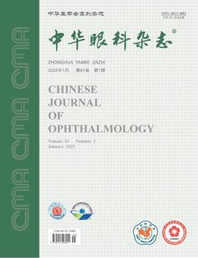
Volume 61 · Issue 01 · 2025
Multilingual International Publishing Platform for Humanities and Social Sciences111
This issue focuses on the research progress of strabismus, amblyopia and pediatric ophthalmology. The responsible editorial committee responsible for the planning, composition and review of this issue is Professor Zhang Wei.
Amblyopia is a common eye disease that seriously affects children′s eye health. The Chinese Association for Pediatric Ophthalmology and Strabismus has carried out a lot of work for the prevention and treatment of amblyopia, but there are still many problems that need to be paid attention to and solved in the amblyopia screening and clinical diagnosis and treatment. In this article, we discuss three aspects of the amblyopia prevention and treatment, including the timing of prevention and treatment according to visual developmental patterns, the standardized treatment of amblyopia, overdiagnosis and commercialization, and the establishment of an efficient amblyopia screening system, for providing guidance for the standardization of clinical practice and improving the level of amblyopia prevention and treatment in China.
Intermittent exotropia (IXT) is the most common type of strabismus, with surgical interventions standing as its main therapeutic modality. In recent years, with the rapid development of the strabismus and pediatric ophthalmology subspecialties, surgical correction for IXT has become a routine practice across numerous institutions in China. However, the surgical success rate is not high and tends to decline with longer follow-up periods. In this article, we highlight key misconceptions and challenges in the surgical management of IXT, with focuses on optimizing the timing of surgical interventions, enhancing the preoperative evaluation, and advancing the standardization of surgical planning, aiming to provide evidence-based recommendations for clinical strategies and improve surgical outcomes of IXT.
Early-onset high myopia (eoHM) in children is a kind of high myopia that occurs in preschool children. While affecting the visual development process, eoHM can cause a series of complications such as retinal rupture and detachment, and posterior scleral staphyloma, which not only damage the visual function, but also delay the overall development of children. Therefore, early prevention, early diagnosis, timely treatment, and long-term management are crucial. This article discusses the necessity and key points of the management of blinding complications caused by eoHM in children, in order to attract doctors′ attention and provide guidance for clinical work.
To explore the efficacy of the modified lateral rectus muscle splitting and nasal transposition surgery in treating large-angle exotropia caused by oculomotor nerve palsy and its impact on ocular motility.
Retrospective case series study. Data was collected from patients diagnosed with large-angle exotropia due to oculomotor nerve palsy and treated by modified lateral rectus muscle splitting and nasal transposition surgery at the Tianjin Eye Hospital from January 2020 to October 2023. The prism and Maddox rod method (for unilateral oculomotor nerve palsy patients) and the Krimsky (prism reflection method) (for bilateral oculomotor nerve palsy patients) were used to measure the degree of strabismus. The adduction of the eye was graded. During the surgery, the lateral rectus muscle was split, and the upper and lower halves were transposed nasally to the upper and lower sides of the medial rectus muscle, respectively. Three patients underwent concurrent medial rectus muscle strengthening surgery. Changes in strabismus degree, ocular motility, equivalent spherical degree, and ocular rotation degree before and after surgery were compared. Surgical success was defined as postoperative alignment within 10△ of orthotropia in primary position by the Krimsky or Hirschberg assessment. Postoperative follow-up was 6 to 12 months.
There were 8 patients (9 eyes) with oculomotor nerve palsy, including 2 females and 6 males, with an age range of 1 to 60 years old. At the end of the follow-up period, 7 patients (7 eyes) had a postoperative strabismus degree within 10△, and 1 patient (2 eyes) was undercorrected. The comparison of the preoperative (-82.78△± 13.94△) and postoperative (-6.22△±8.86△) strabismus degrees in all eyes showed a statistically significant difference (P<0.05). There was no significant difference in the preoperative [(-0.63±1.94) D] and postoperative [(-0.68±1.84) D] equivalent spherical degrees, and the preoperative (3.96°± 13.79°) and postoperative (6.47°±11.96°) rotation degrees (both P>0.05). The preoperative adduction of the eye [-5.0 (-5.0, -4.0)] was significantly different from the postoperative value [-2.0 (-3.5, -2.0)] (P<0.05). Spearman′s correlation analysis showed that the improvement in adduction was not related to the concurrent medial rectus muscle strengthening (r=-0.098, P>0.05), but was related to the preoperative horizontal strabismus degree (r=0.777, P<0.05). No complications such as vision loss, increased intraocular pressure, optic nerve edema, and choroidal detachment occurred in all patients during and after the surgical treatment.
The modified lateral rectus muscle splitting and nasal transposition surgery can effectively and safely correct large-angle exotropia caused by oculomotor nerve palsy and significantly improve the adduction function.
To evaluate the safety and efficacy of cataract extraction combined with multifocal intraocular lens (IOL) optic implantation in Berger space in school-age children with cataracts.
It was a prospective study. The clinical data of school-age children with cataract who underwent cataract extraction combined with multifocal IOL implantation at Qingdao Eye Hospital of Shandong First Medical University from January 2019 to June 2023 were collected. Preoperatively, the examinations of best corrected distance, intermediate and near visual acuity, intraocular pressure, eye position, slit lamp microscopy, and fundus examination were carried out. The optical quality analysis system was used to obtain the objective visual quality indexes such as modulation transfer function cut-off frequency (MTF cut-off), Strehl ratio (SR), and objective scattering index (OSI), and stereopsis was examined. All patients underwent cataract extraction combined with multifocal IOL optic implantation in Berger space. During the follow-up, visual acuity, intraocular pressure, stereopsis, objective visual quality, anterior segment and fundus conditions were examined, and optical coherence tomography (OCT) was used to determine whether there was macular edema and other fundus abnormalities.
A total of 43 cataract patients (86 eyes) were included, with an age of (9.4±3.0) years. Among them, 81 eyes completed cataract extraction combined with multifocal IOL optic implantation in Berger space. The follow-up time was (20.6±13.1) months. During the follow-up, the visual axis remained clear in all eyes. Transient postoperative intraocular hypertension occurred in 5 eyes (25-30 mmHg, 1 mmHg=0.133 kPa), which mostly occurred within 1 week after surgery and could be controlled to the normal range within 2 weeks after drug treatment. During the follow-up, no posterior synechia, secondary glaucoma, IOL decentration, pigmentary IOL deposits, retinal detachment, cystoid macular edema, proliferation of the vitreous anterior boundary membrane, capsular contraction and proliferation, etc. were observed. The preoperative and postoperative best corrected distance visual acuity (logarithm of the minimum angle of resolution) was 0.74±0.56 and 0.07±0.14, respectively, with a statistically significant difference (P<0.001). The best corrected distance visual acuity was≥20/40 in 77 eyes (95.06%), and≥20/29 in 73 eyes (90.12%). The postoperative spectacle independence rate was 77.8% (63/81), and the remaining patients only needed to wear a pair of glasses to correct the reserved or residual refractive error. A total of 5 patients (11.6%) had myopia drift after surgery. The MTF cut-off and SR increased and the OSI decreased after surgery (all P<0.001); the stereopsis was significantly improved (P<0.05).
Multifocal IOL optic implantation in Berger space showed no significant complications and can effectively improve the visual function of school-age children with cataracts. After the correction of distance visual acuity, glasses for near visual acuity are not required.
To investigate the clinical features of Tolosa-Hunt syndrome (THS), a type of painful ophthalmoplegia.
This was a retrospective case series study. The clinical data of patients diagnosed with painful ophthalmoplegia in the Department of Neurology of Tianjin Medical University General Hospital from January 2019 to December 2022 were continuously collected. THS and other types of painful ophthalmoplegia were diagnosed according to the criteria of the International Classification of Headache Disorders, 3rd Edition. The first symptoms, ocular manifestations, imaging and laboratory findings, treatment (glucocorticoids in THS patients) and outcomes were recorded and compared between patients with THS and the other patients.
A total of 35 patients with painful ophthalmoplegia who met the criteria were included. Eighteen patients were diagnosed with THS, including 11 males and 7 females, with an average age of (59.9±13.2) years old (range, 28 to 80 years old). Seventeen patients presented with pain as the initial symptom, 17 patients had ptosis, and 10 patients had abnormal signal lesions in the orbital and cavernous sinus area on MRI on the affected side. Nine patients had abnormal serum immunological results (3 had elevated erythrocyte sedimentation rate and C-reactive protein levels, 6 had positive antinuclear antibodies, and 1 was diagnosed with systemic lupus erythematosus and rheumatoid arthritis). Among the 17 patients with other types of painful ophthalmoplegia, 12 had concurrent diabetes mellitus (including 1 with positive syphilis antibodies and 1 with active pulmonary tuberculosis), 2 had intracranial aneurysms, 1 had ischemic oculomotor nerve palsy, 1 had intracranial infection, and 1 had basal meningitis. Five THS patients experienced symptom relief after 3 days of corticosteroid treatment, and 13 THS patients experienced relief within 1 to 6 weeks of treatment. During the follow-up period (7 to 47 months), 3 patients with THS relapsed.
Most patients with THS have pain as the first symptom, with positive signs of cavernous sinus on MRI and abnormal serum immunologic examination results. Other types of painful ophthalmoplegia usually combine with diabetes mellitus or other systemic diseases, which is helpful in the differential diagnosis.
To observe the characteristics of ocular biological parameters in children with transfusion-dependent β-thalassemia (TDT) and the effect of iron chelator treatment on them.
This was a cross-sectional study. Thirty-two children with TDT (TDT group) and 64 healthy children (control group) who were treated in the Affiliated Hospital of Guangdong Medical University from October 2022 to June 2023 were included. There were 17 males and 15 females in the TDT group, with an age of (10.32±3.58) years; and 34 males and 30 females in the control group, with an age of (10.70±3.23) years. The children with TDT were divided into TDT1 group (using a single iron chelator) and TDT2 group (using two iron chelators in combination) according to the treatment situation of blood transfusion combined with iron chelator. The growth and development indicators such as height, weight and BMI of the children were recorded; the axial length (AL), anterior chamber depth (ACD), lens thickness (LT), central corneal thickness (CCT), corneal horizontal diameter (WTW), and the mean corneal curvature (Km) was calculated; the average size (AVG) of corneal endothelial cells and corneal endothelial cell density (CD) were measured using the corneal endothelium cytometer SP-3000P. to integer values; the blood transfusion time, serum ferritin before and after blood transfusion, the
The height, weight and BMI of the TDT group were all smaller than those of the control group (all P<0.001). The AL of the TDT group was (23.21±0.98) mm, shorter than that of the control group (24.01±1.16) mm; the ACD was (3.49±0.24) mm, shallower than that of the control group (3.63±0.25) mm; the LT was (3.52±0.18) mm, thicker than that of the control group (3.39±0.16) mm; The Km value (44.12±1.59) D is higher than that of the control group (42.93±1.34) D; the CD was (3 035.71±312.59) cells/mm², smaller than that of the control group (3 260.46±248.11) cells/mm² (all P<0.05). In the TDT group, AL and ACD were positively correlated with height (r=0.497,0.454;both P<0.05), and LT was negatively correlated with height (r=-0.400, P<0.05). The age of the TDT2 group was greater than that of the TDT1 group, the LT was smaller than that of the TDT1 group (P>0.05), and the CD was smaller than that of the TDT1 group (P<0.05).
The growth and development of children with TDT are retarded, manifested as short axial length, shallow anterior chamber, thickened lens, higher mean corneal curvature and decreased corneal endothelial cell density. The use of iron chelators may cause corneal changes such as decreased corneal endothelial cell density.
To explore the research status and hotspots in the field of endophthalmitis based on bibliometrics.
A bibliometric study was conducted. In May 2024, the Web of Science Core Collection database was searched for relevant English-language literature on endophthalmitis published from 2014 to 2023. Endnote X9 was used to manage and check the included literature; Excel software was employed for data management and statistics, and graphs were created for frequency data. CiteSpace and VOSviewer software systems were utilized to construct visual maps of literature authors, institutions, countries, journals, and keywords and conduct analyses.
A total of 1 466 articles were finally included. The number of literature in the field of endophthalmitis showed a fluctuating trend in the past decade. The journal with the most published articles was Retina. Among the top 10 authors with the highest number of publications, 5 were from the LV Prasad Eye Institute in India, and 2 were from the Bascom Palmer Eye Institute in the United States. The top 10 institutions with the largest number of literature were the LV Prasad Eye Institute in India, the University of Miami in the United States, Wayne State University in the United States, Aravind Eye Hospital in India, the University of Oklahoma in the United States, Fudan University in China, Manipal International University in Malaysia, Thomas Jefferson University in the United States, Duke University in the United States, and Sun Yat-sen University in China. The United States, India, and China were countries with relatively high productivity in publishing articles on endophthalmitis. Research related to endophthalmitis was dominated by the United States, and the intensity of its international exchanges and cooperation was significantly higher than that of Asian countries, suggesting that other countries, including China, need to strengthen international exchanges. The results of keyword and burst term studies showed that current research on endophthalmitis mainly focused on etiological investigations, risk management, prevention, control, treatment, and prognosis evaluation.
In the past decade, the number of literature in the field of endophthalmitis showed a fluctuating trend. Etiological investigations, incidence rates, and classification management of endophthalmitis were hot research areas, and research reports on prevention, control, diagnosis, treatment, and complex cases were the focus of current and future research.
A 68-year-old male developed foreign body sensation in both eyes, photophobia and tearing in the right eye approximately 6 weeks after initiating dupilumab for severe atopic dermatitis. The right eye presented a crescent-shaped superficial-stromal ulcer in the peripheral cornea with an undermined edge, which was separated from the limbus of the cornea by a clear zone. The left eye had a pannus at the limbus of the cornea. Corneal scraping, bacterial culture, and fungal culture excluded infections. The patient was administered 0.1% fluorometholone eye drops and recombinant bovine basic fibroblast growth factor eye drops. The patient′s condition continued to improve during the follow-up period. At the revisit 82 days after the initial diagnosis, the corneal lesions of the right eye were basically healed, and the corrected visual acuity increased from 0.5 to 1.0.
A 63-year-old male patient presented with a four-month history of vision loss in the left eye and blurred vision in the right eye, the cause of which was not immediately apparent. The patient was initially diagnosed with uveitis in both eyes at the Department of Ophthalmology. Subsequently, the patient was diagnosed as central nervous system lymphoma at the Department of Brain Medicine and initiated a systemic treatment. However, due to the progressive deterioration of visual function, a further evaluation was performed at the Department of Ophthalmology. Based on the results of the vitreous fluid cytokine test, intraocular lymphoma was diagnosed in both eyes. Intravitreal methotrexate injections were administered for eight times and a notable improvement in visual acuity was achieved in both eyes.
The patient was a 62-year-old female. Because of acute painless vision loss in his left eye, he was seen in the Department of Ophthalmology of the First Hospital of Jilin University. Eye examination: corrected visual acuity was 1.0 for the right eye and 0.2 for the left eye; No obvious abnormalities were observed in the anterior segment of both eyes; No obvious abnormalities were observed in the right fundus; The large aneurysm of the supratemporal arch of the retinal vessel in the left eye ruptured, and the bleeding formed a fluid plane, occluding part of the macular area (A in Quintessential Picture 1). Clinical diagnosis; Intra-limiting subretinal hemorrhage of the left eye. 10 min after the left eye was treated with neodymium-doped yttrium aluminum garnet laser, the subretinal hemorrhage in the macular area spread below the vitreous cavity (B in Quintessential Picture 1). Follow-up after treatment, the best corrected visual acuity of left eye was 0.5.
The patient was a 31-year-old female. He went to Hebei Eye Hospital because of painless vision loss in his right eye for 2 days. The patient was previously in good health. Eye examination: visual acuity was 0.1 for the right eye and 1.0 for the left eye; IOP was 13 mmHg (1 mmHg =0.133 kPa) in the right eye and 17 mmHg in the left eye; No obvious abnormalities were observed in the anterior segment of both eyes; A huge off-white round depression on the temporal side of the optic disc of the right eye, with a diameter of 1/2 the diameter of the optic disc, and serous detachment of the retina in the macular area (Quintessential Picture 1). Coherence optical tomography examination: the tissue of the temporal sieve plate of the optic disc of the right eye was missing, and the neuroepithelial detachment of the mastic plaque area was combined (Quintessential Picture 2). Fluorescein fundus angiography examination: the right optic disc fovea showed weak fluorescence in the early stage, gradually increased with time, and became strong fluorescence in the late stage (Essence Picture 3). Clinical diagnosis: Right eye optic disc fovea combined with macular degeneration.
Retinitis pigmentosa (RP) is a group of inherited retinal diseases characterized by progressive loss of photoreceptor cells and retinal pigment epithelium function. Its treatment has long been a focus and challenge in ophthalmic research. Despite advances in therapies such as stem cell transplantation, gene therapy, and retinal prosthetic implants, many difficulties remain. Optogenetic technology, an emerging biological technique combining optics and genetics, holds promise for treating RP by inducing the expression of light-sensitive proteins in degenerated retinal cells, thereby partially restoring patients′ vision and light sensitivity. However, limitations such as difficulties in selecting target cells, potential risks of viral transfection, and uncertainties in visual restoration still exist. With the continuous improvement of light sensitivity of optogenetic proteins and the development of viral vectors with higher transfection efficiency, it is believed that optogenetic therapy will help more patients regain vision.
Immune checkpoint inhibitors (ICI), as a new type of targeted therapeutic drugs, have demonstrated durable efficacy in cancer treatment. However, some patients receiving ICI treatment may be affected by immune-related adverse events (irAE). Compared with irAE in skin tissues, gastrointestinal system, etc., irAE in the neuro-ophthalmic efferent system are relatively rare. The reported irAE in the neuro-ophthalmic efferent system mainly include myasthenia gravis, orbital extraocular myositis, thyroid-associated ophthalmopathy-like conditions, internuclear ophthalmoplegia, Miller-Fisher syndrome, and cranial nerve adverse events. Exploring the possible mechanisms and potential intervention measures of irAE in the neuro-ophthalmic efferent system is crucial for predicting the risk of irAE in patients, formulating preventive measures, and implementing more effective clinical treatments. This review aims to summarize the occurrence mechanisms, clinical features, and prevention and treatment measures of irAE in the neuro-ophthalmic efferent system, so as to assist ophthalmologists and oncologists in the early diagnosis and management of such patients.
CURRENT ISSUE