Multilingual International Publishing Platform for Humanities and Social Sciences111
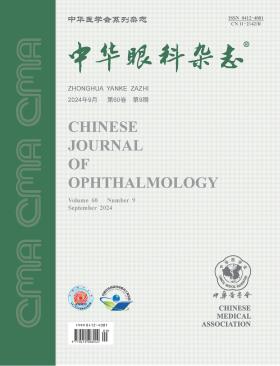
Volume 60 · Issue 09 · 2024
Multilingual International Publishing Platform for Humanities and Social Sciences111
Angle closure is a basic pathological process in primary angle-closure glaucoma. Unlike filtration anti-glaucoma surgery, goniosynechialysis is a treatment for the cause of primary angle-closure glaucoma by reconstructing the physiological aqueous outflow pathway. In the past 10 years, phacoemulsification lens extraction with posterior chamber intraocular lens implantation in combination with goniosynechialysis for the treatment of primary angle-closure glaucoma has become a hot topic of clinical research and discussion. This article points out the issues that need to be paid attention to in the clinical practice of the combined surgery, including the relationship between phacoemulsification surgery and goniosynechialysis, the effectiveness and influencing factors of the combined surgery, and how to avoid the postoperative re-adhesion of the angle, in order to provide reference and guidance for better goniosynechialysis.
Selective laser trabeculoplasty (SLT), as one of the main methods for the treatment of open-angle glaucoma, has again aroused wide concern in recent years. Although SLT has a clear effect on reducing intraocular pressure, its role needs to be fully recognized. In order to provide guidance for the clinical SLT practice, this article summarizes and discusses the current status of SLT treatment for glaucoma, its therapeutic effects on primary open-angle glaucoma, ocular hypertension, and other types of glaucoma, surgical details, and application prospects.
To investigate the anatomical structure changes of the anterior segment and dynamic pupil changes in eyes with suspected primary angle-closure (PACS) under light and dark conditions, and their correlation with the occurrence of acute primary angle-closure (APAC).
This cross-sectional study collected data from 37 PACS patients (66 eyes) who visited the ophthalmology clinic of the Second Affiliated Hospital of Harbin Medical University between September 2019 and March 2021. The patients included 12 males and 25 females, with an average age of (61.27±7.35) years. Of the 66 eyes, 58 had no history of APAC in the contralateral eye, while 8 had a history of APAC in the contralateral eye. Patients without a history of APAC in both eyes underwent the dark room prone provocative test (DRPPT), and the eyes were divided into three groups: DRPPT- (44 eyes), DRPPT+ (14 eyes), and APAC (8 eyes). The DRPPT+ and APAC groups were combined into the APAC+ group. All patients underwent ultrasound biomicroscopy (UBM) to measure the changes in various parameters under light and dark conditions, including anterior chamber angle 500 (ACA500Δ) and 1000 (ACA1000Δ), angle opening distance 500 (AOD500Δ) and 1000 (AOD1000Δ), and iris thickness 500 (IT500Δ) and 1000 (IT1000Δ). Anterior segment analysis was performed to capture the pupil changes under light and dark conditions, recording pupil diameter, maximum dilation speed (Vmax), maximum constriction speed (Vmin), and average speed (Vm).
There was no significant difference in the parameters between DRPPT+ group and APAC group (P>0.05). In the difference analysis, it was found that the Vm value of DRPPT- group [(0.17±0.07) mm/s] was higher than that of APAC+ group [(0.13±0.06)mm/s], and the difference was statistically significant (P<0.05). There was no significant difference in other parameters (P>0.05). Vmax was positively correlated with temporal ACA1000Δ and temporal AOD1000Δ in all patients with PACS (r=0.302, 0.260; P<0.05), Vmin was negatively correlated with temporal ACA1000Δ and temporal AOD1000Δ (r=-0.338, -0.330; P<0.05).
The dynamic changes in the anterior segment and pupil under different lighting conditions provide insights into the risk factors and potential predictive indicators for the occurrence of APAC in PACS patients.
To investigate the factors affecting the progression of visual field defects in patients with myopia and primary open-angle glaucoma (POAG), and to clarify whether the factors vary in patients with different degrees of myopia.
An ambispective cohort study was conducted among patients diagnosed with myopia and POAG from the glaucoma outpatient department at the Zhongshan Ophthalmic Center of Sun Yat-sen University between January 2006 and January 2024. Based on the criteria of functional visual field progression, patients were divided into the progression group and non-progression group, and further divided into the low to moderate myopia subgroup and high myopia subgroup according to the degree of myopia. The patient age, gender, type of glaucoma (high tension glaucoma and normal tension glaucoma), spherical equivalent refraction, best corrected visual acuity (BCVA, recorded as the logarithm of the minimum angle of resolution), intraocular pressure (IOP), central corneal thickness, baseline visual field, history of ophthalmic surgery (corneal refractive surgery and glaucoma surgery), and number of anti-glaucoma medications were summarized. The generalized estimation equation was used for comparison between groups, and the Cox proportional hazards model was used to analyze the factors affecting the progression of visual field defects.
A total of 182 eyes from 106 patients were included in this study. There were 57 eyes in the progression group and 125 eyes in the non-progression group. Compared with the non-progression group, the progression group had the older age [43 (29, 53) years old], worse BCVA [0.05 (0.00, 0.17)], greater IOP fluctuation [1.8 (1.3, 2.9)mmHg(1 mmHg=0.133 kPa)], more common baseline central defects [52.6%(30/57)], higher visual field pattern standard deviations [8.92 (5.32, 12.00)dB], lower visual field index [77% (67%, 88%)], and more anti-glaucoma medications [35.1% (20/57) patients used three medications] (all P<0.05). The Cox proportional hazards models showed that the baseline moderate visual field defects [hazard ratio (HR)=2.33, 95% confidence interval (CI): 1.25 to 4.36, P=0.008], baseline central defects (HR=2.09, 95%CI: 1.11 to 3.93, P=0.022), older age (Model A, HR=1.03, 95%CI: 1.00 to 1.05, P=0.017; Model B, HR=1.02, 95%CI: 1.00 to 1.05, P=0.019), and greater IOP fluctuation (Model A, HR=1.54, 95%CI: 1.32 to 1.81, P<0.001; Model B, HR=1.49, 95%CI: 1.26 to 1.75, P<0.001) were risk factors for visual field progression. In the low to moderate myopia subgroup, the increased risk of progression was associated with baseline central defects (HR=5.74, 95%CI: 1.72 to 19.20, P=0.005), worse BCVA (Model A, HR=15.80, 95%CI: 2.07 to 121.00, P=0.008; Model B, HR=12.50, 95%CI: 2.65 to 58.70, P=0.001), and older age (Model A, HR=1.05, 95%CI: 1.02 to 1.08, P=0.002; Model B, HR=1.07, 95%CI: 1.03 to 1.11, P<0.001). In the high myopia subgroup, the increased risk of progression was associated with baseline moderate visual field defects (HR=2.35, 95%CI: 1.12 to 4.92, P=0.024) and greater IOP fluctuation (Model A, HR=1.50, 95%CI: 1.24 to 1.82, P<0.001; Model B, HR=1.52, 95%CI: 1.26 to 1.83, P<0.001).
Age, IOP fluctuation, baseline moderate visual field defects, and baseline central defects were the factors affecting the progression of visual field defects in patients with myopia and POAG. There were differences in the influencing factors of visual field progression in patients with different degrees of myopia.
To evaluate the efficacy and safety of internal XEN gel stent implantation or combined with phacoemusification and intraocular lens (IOL) implantation in the treatment of glaucoma.
It was a retrospective case series study. Forty-five patients (28 males and 17 females; 52 eyes) who had different types of glaucoma treated at the Shenzhen Eye Hospital from December 2021 to June 2023 were included. XEN gel stent implantation (the XEN group) or XEN implantation combined with phacoemusification and IOL implantation (the combined group) were performed in these patients. The postoperative follow-up was 18 months. The observational indexes included the best corrected visual acuity (recorded as the logarithm of the minimum angle of resolution), intraocular pressure (IOP), XEN implantation quadrant, number of IOP-lowering medications, corneal endothelial cell count, operation success rate and surgical complications. The one-way repeated measures of variance, LSD-t test, and rank sum test were used for statistical analysis.
There were 28 patients (33 eyes) in the XEN group and 17 patients (19 eyes) in the combined group. The postoperative best corrected visual acuity had no significant change in the XEN group, but improved significantly (0.21±0.30 at 6 months; P<0.05) in the combined group, compared with the preoperative value. The IOP of the two groups at 18 months after surgery [(12.2±3.1) and (11.9±3.9) mmHg (1 mmHg=0.133 kPa)] was significantly lower than that before surgery [(22.1±8.5) and (19.4±10.2) mmHg; P<0.05]. The average number of IOP-lowering medications in all patients was 3 (2, 3) before surgery and 0 (0, 0) at 18 months after surgery. The difference was statistically significant (P<0.05). In the XEN group, the corneal endothelial cell count was (2 387.37±478.22) cells/mm² preoperatively and (2 193.89±311.96) cells/mm² at 12 months. The decrease showed no statistical significance. The XEN gel stent was implanted in the supranasal quadrant in 8 eyes (15.4%), in the inferonasal quadrant in 36 eyes (69.2%), and in the infratemporal quadrant in 8 eyes (15.4%). The operation success rate [complete success rate, 86.5% (45/52); conditional success rate, 9.6% (5/52)] was 96.2% (50/52). The filtering bleb scarring occurred in 9.6% (5/52) of eyes. A small amount of hyphema, postoperative shallow anterior chamber, transient hypertension, and intraoperative drain breakage were found in 7.7% (4/52) of eyes, respectively. A small amount of subconjunctival bleeding was observed in 5.8%(3/52) of eyes. Re-implantation of an XEN gel stent was performed in 5.8% (3/52) of eyes. The intraoperative conjunctival tissue rupture, hypotony macular edema, and drain exposure were observed in 1.9% (1/52) of eyes, respectively.
The XEN gel stent implantation or combined with phacoemusification and IOL implantation was effective and safe in treating different types of glaucoma, but its long-term clinical efficacy needs further follow-up observations in multi-center, larger-scale research.
To explore the clinical phenotypes and pathogenic gene variation characteristics of three Chinese Han ethnic families affected by Nance-Horan syndrome, a rare X-linked genetic disorder.
A pedigree investigation study was conducted at the First Affiliated Hospital of Zhengzhou University, collecting clinical data from three Chinese Han families with Nance-Horan syndrome between February 2009 and September 2018. Detailed family histories, comprehensive ophthalmological and systemic examinations were documented. Pedigree charts were created, and genetic inheritance patterns were analyzed to preliminarily diagnose the probands and other affected individuals. Genomic DNA was extracted from peripheral blood samples of family members, and next-generation sequencing was used to screen for target gene variations, which were confirmed by Sanger sequencing. Pathogenicity of the genetic variants and their impact on three-dimensional protein structure were analyzed using MutationTaster and computer-aided protein modeling.
In Family 1, there are 5 patients, including 4 females (aged 42, 37, 9 and 7) and 1 males (aged 12). In Family 2, there are 5 patients, including 3 females (aged 54, 32 and 16) and 2 males (aged 26 and 9). In Family 3, there are 8 patients, including 5 females (aged 69, 42, 37, 35 and 14) and 3 males (aged 10, 7 and 4). All probands in the three families exhibited nuclear cataracts with typical congenital hereditary cataract features, but no noticeable abnormalities in facial appearance or teeth. Next-generation sequencing identified new variation sites in the NHS gene, specifically c.2519_2520del, exon3del, and c.3847C>T. These variations included nonsense mutation p.(Ser840*), exon deletion p.(?), and nonsense mutation p.(Gln1283*). Combined clinical and genetic sequencing results confirmed X-linked Nance-Horan syndrome in all three families. Bioinformatics analysis indicated these variation sites were pathogenic and resulted in abnormal three-dimensional protein structures, likely being the main cause of Nance-Horan syndrome.
The majority of patients from the three Nance-Horan syndrome families studied were affected by congenital hereditary cataracts characterized by nuclear opacities.The NHS gene variations c.2519_2520del, exon3del, and c.3847C>T are newly identified pathogenic sites in Nance-Horan syndrome, reported for the first time across three different families.
To investigate the action potential firing patterns of neurons in the visual sensory layers of the superior colliculus in early postnatal mice and the electrophysiological characteristics of neurons with different firing patterns.
This experimental study utilized whole-cell patch-clamp recordings performed on neurons in the visual sensory layers of the superior colliculus using brain slices from 57 healthy male C57BL/6J mice aged 14 to 20 days (weighing 5.0 to 8.9 g) using brain slices. In current-clamp mode, action potential characteristics were analyzed based on the first action potential generated by depolarizing current, and the firing patterns of neurons were recorded using step depolarizing currents. Neuronal firing patterns were analyzed using hierarchical clustering, and the active electrical properties of neurons with different firing patterns were compared.
A total of 135 neurons from the visual sensory layers of the superior colliculus were successfully recorded. Cluster analysis of the neuronal firing patterns identified three types of firing patterns: tonic firing (97, 72%), phasic firing (26, 19%), and single firing (12, 9%). The number of action potentials for each firing pattern was 13.30±7.38, 3.73±3.61, and 0.83±0.39, respectively, with significant differences (P<0.001). There was no significant difference in the membrane potential response to step currents among the three firing pattern types (P>0.05). The action potential amplitudes were (60.45±12.22), (53.67±13.20), and (44.04± 12.92) mV, and the afterhyperpolarization amplitudes were (13.45±13.79), (12.02±13.11), and (20.75±2.85) mV, respectively. The maximum rising slopes were (171.29±77.46), (130.14±61.83), and (78.89±37.08) V/s, and the maximum falling slopes were (-76.33±33.61), (-68.17±31.65), and (-47.97±13.92) V/s, respectively, with all differences being statistically significant (all P<0.05). There were no significant differences in the resting membrane potential, action potential threshold, half-width, and afterhyperpolarization duration among the three firing pattern types (all P>0.05).
In the early postnatal mice, neurons in the visual sensory layers of the superior colliculus exhibit three distinct firing patterns: tonic, phasic, and single firing. These firing pattern types show significant differences in action potential amplitude, afterhyperpolarization amplitude, maximum rising slopes, and maximum falling slopes.
The patient is a 28-year-old male who presented with hemifield slide for 10 months following surgery for a large pituitary adenoma. A comprehensive ophthalmological examination and visual function tests were conducted, including best corrected visual acuity, visual field, stereopsis, accommodative convergence, Worth four-dot test, and prism cover test for quantifying strabismus, along with assessments using a depression scale and a visual function questionnaire. Rehabilitation plans included applying press-on prisms to correct vertical strabismus, binocular vision training, and saccadic training. After rehabilitation, the patient was able to maintain binocular single vision at near and intermediate distances, experienced improved visual comfort, and showed significant improvements in scores on the depression scale and visual function questionnaire.
The patient is a 2-year-old male. The family consulted the Department of Ophthalmology, Shanghai Ninth People′s Hospital, after noticing a white reflection in the pupil area of the child′s right eye for 6 days. Following a thorough ocular and systemic examination, the patient was diagnosed with retinoblastoma (Group E, cT2bN0M0) of the right eye. The right eye was enucleated and classified as pathological stage pT3cN0M0. Postoperatively, systemic intravenous chemotherapy with the VEC regimen was administered. Genetic testing revealed a germline mutation in the RB1 gene: c.874 (exon9) delT (p.Tyr292fsTer9), necessitating close monitoring of the socket during follow-up visits. Three months after the operation, fundus examination revealed yellow-white lesions in the left eye, and bilateral retinoblastoma was diagnosed (Group E in the right eye, Group C in the left eye). Based on the ICRB and pTNM stages, the patient underwent six rounds of systemic intravenous chemotherapy and three rounds of cryotherapy in the left eye. No recurrence was detected with a 4-year follow-up. The patient was initially diagnosed with unilateral retinoblastoma, but later developed the disease in the contralateral eye during treatment, which was a case of metachronous bilateral retinoblastoma.
The patient was a 42-year-old female. Because of blurred vision in his left eye for 1 month, he went to the Department of Ophthalmology, the First Hospital of Jilin University. History of ocular trauma, surgery, contact lens wear and systemic disease were denied. Eye examination: the corrected visual acuity of both eyes was 1.0; IOP was 15.0 mmHg (1 mmHg =0.133 kPa) in the right eye and 14.0 mmHg in the left eye; The corneal epithelial cells above the nose of the left eye are striped gray-white and turbid, extending from the limbus above the nose to the central pupil area, resembling a phoenix, with a lesion range of 64.14 mm ×16.17 mm, and a clear boundary with the normal cornea (Quintessence Picture 1); The cornea of the right eye is transparent; The anterior chamber of both eyes was clear, without retrocorneal deposits, and no obvious abnormalities in iris and lens were observed; No obvious abnormalities were found in the fundus of both eyes. Anterior segment coherence light tomography examination: A flaky hyperreflective signal was seen in the corneal epithelial cell layer of the left eye, with uniform brightness and density, and the depth of the lesion was limited to the corneal epithelial layer (Quintessential Picture 2). Corneal confocal microscopy: The cytoplasm of the corneal epithelial pterygoid cell layer in the lesion area of the left eye was highly reflective, surrounding the hyporeflective nucleus, and clearly bounded with the normal epithelial area. Clinical diagnosis: Lisch's corneal dystrophy in the left eye.
The regulation of blood flow in the human eye, aqueous humor production, and the outflow channels of aqueous humor are all controlled by the autonomic nervous system (ANS), encompassing both sympathetic and parasympathetic nerves. Patients with primary glaucoma often exhibit autonomic nervous system dysfunctions, which may exacerbate visual impairment. The homeostasis of the autonomic nervous system is influenced by circadian rhythms, physical exercise, emotional states, medications, and other factors. Previous studies have indicated that activities such as aerobic exercise, yoga breathing, and meditation can promote the restoration of autonomic nervous system balance, thereby reducing intraocular pressure. However, further evidence is required to substantiate the efficacy of ANS activity regulation as an effective adjunct therapy for primary glaucoma. This review examines the role and mechanisms of autonomic nervous system regulation in the context of primary glaucoma.
Neovascular retinal diseases pose a significant burden, often resulting in visual impairment. Intravitreal injection of anti-vascular endothelial growth factor (VEGF) drugs serves as the primary therapeutic approach. Nonetheless, certain patients necessitate continued anti-VEGF treatment post-vitrectomy or other ocular surgeries. Emerging evidence suggests that variations in surgical techniques and postoperative vitreous cavity management may induce distinct intraocular pharmacokinetics (PK) of anti-VEGF agents following vitrectomy, prompting potential adjustments in therapeutic strategies. This review offers a thorough examination of the pharmacokinetic determinants impacting anti-VEGF drugs and their intraocular dynamics post-vitrectomy.
Primary angle closure glaucoma (PACG) is a blinding eye disease with high incidence. Lens factors play an important role in the occurrence and development of glaucoma. In this paper, the pathogenesis and evaluation indexes of lens thickness, relative position and morphology, abnormal suspensory ligament and incomplete dislocation of lens, ciliary body block and translens pressure difference in the occurrence and development of PACG were analyzed, and the related research results of relieving lens factors in the treatment of PACG were summarized, so as to provide references for predicting the progression of PACG and guiding clinical treatment.
CURRENT ISSUE