Multilingual International Publishing Platform for Humanities and Social Sciences111
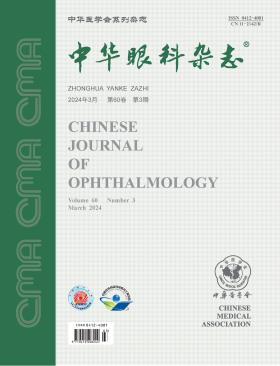
Volume 60 · Issue 03 · 2024
Multilingual International Publishing Platform for Humanities and Social Sciences111
This issue focuses on the research progress in the diagnosis and treatment of fundus diseases. The responsible editorial committee responsible for the planning, composition and review of this issue is Professor Wei Wenbin.
Myopic maculopathy is the primary cause of irreversible visual impairment in patients with pathologic myopia, and myopic traction maculopathy often requires vitrectomy for treatment. Myopic traction maculopathy encompasses epiretinal membrane, foveoschisis, macular hole, and macular hole-related retinal detachment. It is recommended to perform vitrectomy combined with inner limiting membrane peeling for Type II epiretinal membrane, foveal-sparing inner limiting membrane peeling for foveoschisis, inverted inner limiting membrane flap technique for macular hole, and vitrectomy combined with macular buckle for refractory macular hole-related retinal detachment. Myopic traction maculopathy is a chronically progressive condition, and surgeons need to accurately determine the timing of surgery and choose appropriate procedures to maximize the benefits for patients.
Neovascular age-related macular degeneration (nAMD) is a leading cause of blindness in the elderly, and anti-vascular endothelial growth factor (VEGF) therapy is currently the primary treatment approach. However, the real-world effectiveness of nAMD treatment is not always satisfactory and faces various challenges. Frequent administration and follow-up burdens can lead to decreased patient compliance during long-term treatment, resulting in suboptimal outcomes. Some lesions exhibit poor or no response to anti-VEGF treatment, leading to difficulties in maintaining or even declining visual acuity. Factors such as lesion fibrosis and tissue atrophy can contribute to visual deterioration. Therefore, standardizing and individualizing treatment plans, along with enhancing comprehensive monitoring and management throughout the disease course, are crucial improvement measures. The evidence-based guidelines for diagnosis and treatment of age-related macular degeneration in China, released in 2023, provide guidance for standardized clinical diagnosis and treatment. Meanwhile, research and development of new drugs and administration methods are anticipated for the future.
Internal limiting membrane (ILM) peeling is a critical step in the process of macular hole surgery, giving rise to various modified techniques such as ILM flip-over coverage, ILM and other tissue tamponade procedures, and foveal-sparing ILM peeling. All these approaches aim to improve the postoperative closure rate of macular holes. The goal of macular hole surgery is to better preserve the integrity of the foveal center structure, with the aim of achieving functional recovery on the basis of anatomical restoration. However, in clinical practice, there is a tendency to excessively choose certain surgical methods solely to pursue the closure rate of the hole, which may not be beneficial for the visual function recovery of the patients. This article discusses how to correctly select the internal limiting membrane and its derivative procedures in macular hole surgery, combining clinical practice and relevant domestic and international research literature. It aims to provide insights for colleagues performing macular hole surgery as a reference regarding this clinical focus issue.
Autosomal dominant optic atrophy (ADOA) primarily affects retinal ganglion cells and their axons, resulting in varying degrees of central vision loss from childhood. Due to the rarity of ADOA in clinical practice, Chinese ophthalmologists currently lack sufficient understanding of the disease and experience non-standardized diagnostic procedures and high clinical and genetic misdiagnosis rates. To address these issues, the Ophthalmology Group of China Alliance for Rare Diseases/Beijing Society of Rare Disease Clinical Care and Accessibility and the Neuro-ophthalmology Group of Ophthalmology Branch of Chinese Medical Association have established an expert panel to form consensus opinions based on extensive discussions. This consensus would enhance the knowledge and diagnostic capabilities of Chinese clinicians regarding ADOA and promote awareness of related treatment principles and genetic counseling.
To investigate the efficacy of pars plana vitrectomy (PPV) without intraocular tamponade in the treatment of high myopic eyes with myopic foveoschisis (MF) accompanied by foveal detachment (FD).
A retrospective case series study was conducted. The medical records of patients diagnosed with unilateral MF accompanied by FD at the Eye & ENT Hospital of Fudan University between May 2018 and December 2021 were collected. All patients underwent 23-gauge PPV with posterior vitreous cortex clearance, and no intraocular tamponade was applied. The cases were divided into groups based on whether the internal limiting membrane was peeled during surgery or retained. Follow-up was conducted for at least 12 months. The main outcome measures included postoperative best-corrected visual acuity (BCVA, converted to logarithm of the minimum angle of resolution), central foveal thickness (CFT), MF resolution, and complications. Statistical analyses were performed using t-tests, chi-square tests, Fisher′s exact tests, and univariate and multivariate linear regression.
A total of 40 patients (40 eyes) with MF and FD were included in the study, with 30.0% being male and 70.0% female. The mean age was (56.9±11.7) years, and the axial length of the eyes was (29.1±1.9) mm. At 12 months postoperatively, BCVA improved from baseline 1.15±0.58 to 0.73±0.39 (t=6.11, P<0.001), and CFT decreased from baseline (610.1±207.2) μm to (155.9±104.1) μm (t=13.47, P<0.001). Complete resolution of MF with foveal reattachment was observed in 80.0% of eyes, with a median time of 6 (5, 8) months. There was no significant difference in BCVA and CFT between the internal limiting membrane peeled group and retained group [0.68±0.39 vs. 0.79±0.40, t=0.85, P=0.403; (148.3±63.8)vs.(164.3±137.2)um,t=0.48, P=0.634]. One eye experienced macular hole and another eye developed retinal detachment postoperatively. Correlation analysis showed a positive correlation between BCVA at 12 months postoperatively and baseline BCVA (β=0.433, P<0.001).
Pars plana vitrectomy without intraocular tamponade is effective in treating MF accompanied by FD. The choice between internal limiting membrane peeling and retention does not significantly affect visual prognosis.
To utilize a Python-based fluorescence area detection system to observe and quantitatively analyze the intraocular distribution characteristics and metabolic patterns of Indocyanine Green (ICG) following epiretinal membrane peeling.
A prospective case series study was conducted on patients with idiopathic epiretinal membrane undergoing vitrectomy at West China Hospital of Sichuan University from March 2019 to March 2021. ICG staining was applied during surgery for peeling the epiretinal membrane and internal limiting membrane. Patients were followed up at 1 week, 1 month, 3 months, 6 months, and 12 months postoperatively, with assessments including best-corrected visual acuity, intraocular pressure, fundus photography, near-infrared fundus fluorescence imaging (NIR-FF), and optical coherence tomography (OCT). A Python-based ICG intraocular metabolism detection system was developed to measure the residual area of ICG fluorescence on NIR-FF, predict the ICG metabolic pattern equation, and correlate it with postoperative visual acuity and peripapillary retinal nerve fiber layer thickness.
A total of 64 patients (64 eyes) were included, with an average age of 64.6±8.4 years, including 25 males (39.1%) and 39 females (60.9%). Preoperative NIR-FF images showed no ICG strong fluorescence. At 1 week postoperatively, diffuse ICG strong fluorescence appeared in the posterior pole, and the internal limiting membrane removal area exhibited a ring-like weak fluorescence. Over time, ICG strong fluorescence was observed along the vascular arch and nerve fiber trajectory, gradually diminishing toward the optic disc, with residual ICG fluorescence still visible at the optic disc at 1 year. The Python-based ICG fluorescence area detection system effectively measured intraocular residual ICG area. A predictive equation for the 12-month residual ICG area was constructed through linear regression analysis (Residual ICG area=0.22 × Residual ICG area at 6 months, R2=16%, P=0.002). Except for a negative correlation between the ICG residual area at 1 month and postoperative visual acuity (P=0.017, r=-0.195), no correlation was found between intraocular ICG fluorescence residual area and postoperative visual acuity or peripapillary retinal nerve fiber layer thickness at other follow-up times (all P>0.05).
In patients with idiopathic epiretinal membrane undergoing ICG staining for internal limiting membrane peeling, ICG exhibits characteristic metabolic processes in the eye, with strong fluorescence along the vascular arch and nerve fiber trajectory, gradually converging toward the optic disc over time. The Python-based ICG fluorescence area detection system provides a clear display of the intraocular distribution characteristics of ICG after epiretinal membrane peeling and serves as a tool for predicting the metabolic patterns of ICG in the eye.
To analyze the changes in optical coherence tomography angiography(OCTA)-related parameters before and after idiopathic macular hole (IMH) internal limiting membrane (ILM) flap inversion surgery and assess their impact on retinal sensitivity.
A retrospective case series study was conducted, collecting clinical data of 30 patients (30 eyes) diagnosed with IMH who underwent vitrectomy combined with ILM flap inversion surgery at Shanxi Eye Hospital, affiliated with Shanxi Medical University, between January 2020 and December 2021. Visual acuity and best-corrected visual acuity were examined preoperatively and at 1, 3, and 6 months postoperatively. Microperimetry measured retinal sensitivity (RS), and OCTA measured retinal thickness (RT) as well as vessel density (VD) in the superficial capillary plexus (SCP) and deep capillary plexus (DCP). Statistical analysis was performed using t-tests, repeated measures analysis of variance, and Pearson correlation analysis.
Thirty patients with IMH (30 eyes) were included, with 3 males (3 eyes) and 27 females (27 eyes). The mean age was (62.5±3.0) years, and the follow-up time was (96.3±1.3) days, with a 100% closure rate of macular holes postoperatively. RT in the macular was significantly lower at 1, 3, and 6 months postoperatively compared to preoperative values (F=46.21, P<0.001). The RT in the upper macular region showed statistically significant differences at different time points (P<0.001). VD in the SCP layer showed no significant differences between the upper and lower macular regions at various time points (F=3.21, P=0.601). VD in the upper region of the DCP layer increased at 1, 3, and 6 months postoperatively (P<0.001). RS in the macular was higher at 1, 3, and 6 months postoperatively compared to preoperative values (F=52.01, P<0.001). RS in the lower macular region increased at 3 and 6 months postoperatively (P<0.001), while in the upper region, it increased only at 6 months postoperatively (P<0.001). There was a positive correlation between RS and RT at 1 and 3 months postoperatively, but not at 6 months postoperatively in the upper macular region (r=0.40, P=0.071). In the lower macular region, there was a positive correlation between RS and RT at 1 and 3 months postoperatively (P<0.001). There was no correlation between RS in the upper macular region at 6 months postoperatively and preoperative RT (r=0.43, P=0.072), but there was a positive correlation with RT at 3 months postoperatively (r=0.58, P=0.041).
After idiopathic macular hole internal limiting membrane flap inversion surgery, the OCTA-related parameters have changed. There are transient changes in deep vascular parameters and thinning of the retinal layers at the ILM inversion site, leading to decreased sensitivity.
To achieve automatic segmentation, quantification, and grading of different regions of leopard spots fundus (FT) using deep learning technology. The analysis includes exploring the correlation between novel quantitative indicators, leopard spot fundus grades, and various systemic and ocular parameters.
This was a cross-sectional study. The data were sourced from the Beijing Eye Study, a population-based longitudinal study. In 2001, a group of individuals aged 40 and above were surveyed in five urban communities in Haidian District and three rural communities in Daxing District of Beijing. A follow-up was conducted in 2011. This study included individuals aged 50 and above who participated in the second 5-year follow-up in 2011, considering only the data from the right eye. Color fundus images centered on the macula of the right eye were input into the leopard spot segmentation model and macular detection network. Using the macular center as the origin, with inner circle diameters of 1 mm, 3 mm, and outer circle diameter of 6 mm, fine segmentation of the fundus was achieved. This allowed the calculation of the leopard spot density (FTD) and leopard spot grade for each region. Further analyses of the differences in ocular and systemic parameters among different regions′ FTD and leopard spot grades were conducted. The participants were categorized into three refractive types based on equivalent spherical power (SE): myopia (SE<-0.25 D), emmetropia (-0.25 D≤SE≤0.25 D), and hyperopia (SE>0.25 D). Based on axial length, the participants were divided into groups with axial length<24 mm, 24-26 mm, and>26 mm for the analysis of different types of FTD. Statistical analyses were performed using one-way analysis of variance, Kruskal-Wallis test, Bonferroni test, and Spearman correlation analysis.
The study included 3 369 participants (3 369 eyes) with an average age of (63.9±10.6) years; among them, 1 886 were female (56.0%) and 1, 483 were male (64.0%). The overall FTD for all eyes was 0.060 (0.016, 0.163); inner circle FTD was 0.000 (0.000, 0.025); middle circle FTD was 0.030 (0.000, 0.130); outer circle FTD was 0.055 (0.009, 0.171). The results of the univariate analysis indicated that FTD in various regions was correlated with axial length (overall: r=0.38, P<0.001; inner circle: r=0.31, P<0.001; middle circle: r=0.36, P<0.001; outer circle: r=0.39, P<0.001), subfoveal choroidal thickness (SFCT) (overall: r=-0.69, P<0.001; inner circle: r=-0.57, P<0.001; middle circle: r=-0.68, P<0.001; outer circle: r=-0.72, P<0.001), age (overall: r=0.34, P<0.001; inner circle: r=0.30, P<0.001; middle circle: r=0.31, P<0.001; outer circle: r=0.35, P<0.001), gender (overall: r=-0.11, P<0.001; inner circle: r=-0.04, P<0.001; middle circle: r=-0.07, P<0.001; outer circle: r=-0.11, P<0.001), SE (overall: r=-0.20; P<0.001; inner circle: r=-0.19, P<0.001; middle circle: r=-0.20, P<0.001; outer circle: r=-0.20, P<0.001), uncorrected visual acuity (overall: r=-0.18, P<0.001; inner circle: r=-0.26, P<0.001; middle circle: r=-0.24, P<0.001; outer circle: r=-0.22, P<0.001), and body mass index (BMI) (overall: r=-0.11, P<0.001; inner circle: r=-0.13, P<0.001; middle circle: r=-0.14, P<0.001; outer circle: r=-0.13, P<0.001). Further multivariate analysis results indicated that different region FTD was correlated with axial length (overall: β=0.020, P<0.001; inner circle: β=-0.022, P<0.001; middle circle: β=0.027, P<0.001; outer circle: β=0.022, P<0.001), SFCT (overall: β=-0.001, P<0.001; inner circle: β=-0.001, P<0.001; middle circle: β=-0.001, P<0.001; outer circle: β=-0.001, P<0.001), and age (overall: β=0.002, P<0.001; inner circle: β=0.001, P<0.001; middle circle: β=0.002, P<0.001; outer circle: β=0.002, P<0.001). The distribution of overall (H=56.76, P<0.001), inner circle (H=72.22, P<0.001), middle circle (H=75.83, P<0.001), and outer circle (H=70.34, P<0.001) FTD differed significantly among different refractive types. The distribution of overall (H=373.15, P<0.001), inner circle (H=367.67, P<0.001), middle circle (H=389.14, P<0.001), and outer circle (H=386.89, P<0.001) FTD differed significantly among different axial length groups. Furthermore, comparing various levels of FTD with systemic and ocular parameters, significant differences were found in axial length (F=142.85, P<0.001) and SFCT (F=530.46, P<0.001).
The use of deep learning technology enables automatic segmentation and quantification of different regions of theFT, as well as preliminary grading. Different region FTD is significantly correlated with axial length, SFCT, and age. Individuals with older age, myopia, and longer axial length tend to have higher FTD and more advanced FT grades.
To evaluate the effects of re-tucking the superior oblique muscle on recurrent or residual compensatory head position.
A retrospective case series study was conducted. 12 recurrent or residual compensatory head position patients (12 eyes) with congenital superior oblique palsy who underwent superior oblique re-tucking in Tianjin Eye Hospital from March 2015 to February 2021 were included. All patients had a history of superior oblique tucking procedure and showed signs of superior oblique muscle palsy without inferior oblique muscle overaction. During surgery, the Guyton forced duction test is used to evaluate the relaxation of the superior oblique muscle tendon, which affects the re-tucking length of the muscle.Their head position, vertical deviation, eye movement, fovea-disa angle, and Bielschowsky head tilt test were assessed pre-and post-surgery. Statistical analysis was performed using ttest and paired samples Wilcoxon signed rank test.
Out of the 12 patients, 8 were male and 4 were female, aged between 2 and 9 years. The initial surgery was done at age 6, with a superior oblique recession length of (7.17±1.03) mm. Recurrent head tilt occurred in 11 patients after (3.82±0.98) months postoperatively, and 1 patient had residual head tilt, with a followup period of six months or more. Ocular motility examination revealed underaction of the superior oblique muscle, positive Bielschowsky′s head tilt test, and Guyton forced duction tese indicating relaxation of the paralyzed superior oblique muscle tendon. Scar adhesion was observed at the stop of the superior oblique muscle, as well as the previous sutures. The scar and the sutures around the stop of the superior oblique muscle were released, the mean re-tucking amount was(7.83±1.59)mm. Follow-up at 12 to 18 months postoperatively showed disappearance of compensatory head position, significant improvement in superior oblique muscle lag, normal ocular motility, and no occurrence of Brown syndrome. The results of Bielschowsky head tilt were negative in 9 cases and still positive in 3 cases after superior oblique re-tucking. The primary vertical deviation was 2.5 (2.0, 5.3) prism diopter pre-operatively and 1 (0, 1) prism diopter post-operatively, respectively. The difference was statistically significant (U=6.00, P<0.001). The total amount of FDA in both eyes was (-22.04±5.47)° and (-15.27±6.08)° pre-and post-operatively, respectively. The difference was statistically significant (t=2.87, P=0.009). All 12 patients have normal eye movement after superior oblique re-tucking procedure. All patients had no compensatory head position at last follow-up.
Superior oblique re-tucking is suitable for patients with relaxation of the superior oblique muscle tendon and extrocular rotation as the main sign. It can effectively and safely correct the recurrent or residual compensatory head position after re-tucking the superior oblique muscle.
A 61-year-old male patient presented with blurred vision in the right eye for 1 day. The patient had previously undergone phacoemulsification with intraocular lens implantation (10 years ago) and intravitreal implantation of dexamethasone (due to uveitis) in the eye. There was edema in the inferior cornea, along with Descemet membrane folds. The rod-shaped dexamethasone implant was visible in the inferior anterior chamber. Without pupil dilation, the patient was asked to keep a supine position and avoid head tilting for 1 day. The implant spontaneously relocated into the vitreous cavity, resulting in a reduction of corneal edema. This suggests that the dislocation of the intravitreal implant into the anterior chamber may be caused by a local zonular abnormality, and the dislocated implant has the potential to reposition itself spontaneously.
A 31-year-old female patient with refractive error in both eyes underwent small incision lenticule extraction. On the 4th day after surgery, arc-shaped peripheral corneal infiltrates appeared in the right eye. Tobramycin and dexamethasone eye drops, 0.3% gatifloxacin eye drops, and a corneal bandage lens were applied to the eye. After bacterial infection was ruled out, dexamethasone sodium phosphate was injected subconjunctivally near the corneal lesion. The symptoms improved and the corneal lesion subsided afterwards.
An 81-year-old female patient experienced a rapid increase in the volume of a rice-sized black mass on the left eye over a period of six months. The mass extended out of the eye and exhibited surface erosion with accompanying hemorrhage. Following a live tissue examination and histopathology after orbital exenteration under general anesthesia, the diagnosis of a giant malignant palpebral conjunctival melanoma of the spindle cell subtype was confirmed. The patient refused to undergo local radiation therapy or systemic chemotherapy and died from the disease six months later.
The patient was a 52-year-old male. Because the visual acuity of his right eye decreased for 3 days, he went to the ophthalmology department of Liuzhou Workers' Hospital. Deny history of systemic diseases such as hypertension and diabetes. Eye examination: visual acuity was 0.5 for the right eye and 1.0 for the left eye; IOP was 15 mmHg (1 mmHg =0.133 kPa) in the right eye and 17 mmHg in the left eye; No obvious abnormalities were observed in the anterior segment of both eyes; Dendritic retinal abnormal vascular anastomosis involving the macula with tortuous dilatation of the subtemporal retina of the right fundus (Quintessential Picture 1). Fluorescein fundus angiography examination: Abnormal subtemporal retinal vessels in the right ophthalmic artery phase are partially filled with obvious stratification; Early subtemporal abnormal retinal vessels are completely filled, while the normal superior temporal vein begins to fill, but the normal inferior temporal vein is not yet filled (Quintessence Picture 2); Late no fluorescein leakage. Coherence light tomography examination: abnormal vascular signals in the macular neuroepithelial layer of the right eye, foveal neuroepithelial layer detachment (Quintessential Picture 3). Clinical diagnosis: congenital retinal vascular abnormality in right eye; Central serous chorioretinopathy of the right eye.
Thyroid-associated ophthalmopathy (TAO) is an autoimmune eye disease that affects visual function and appearance, involving pathological remodeling processes of orbital tissue such as inflammatory reaction, oxidative stress, lipogenesis, and fibrosis. Current clinical first-line treatment options cannot be effective for all patients. This article summarizes the research on potential therapeutic targets of TAO at home and abroad in recent years, including receptor protein targets, immune cell targets, fat suppression targets, anti-fibrosis targets, transcription factor targets, and metabolic regulatory enzyme targets. Both non-natural compounds and natural compounds are introduced, with a view to providing clinical researchers with reference and ideas in the treatment of TAO and promoting the clinical application of new therapeutic drugs.
Adeno-associated virus (AAV) vectors have been widely employed in gene therapy for ocular and systemic diseases. However, clinical trial outcomes have indicated that gene therapy may trigger severe adverse events associated with immune-inflammatory reactions, thereby impacting the safety and efficacy of gene therapy. The immune-inflammatory reaction induced after gene therapy in the eye is referred to as gene therapy-associated uveitis, which has become a major obstacle limiting the long-term and effective use of ocular gene therapy. This review comprehensively explores four aspects: the immune response mechanisms of gene therapy, ocular manifestations of associated uveitis, factors influencing immune inflammation, and preventive and therapeutic strategies. The aim is to provide insights for the development of safer and more effective ocular gene therapy.
Sleep apnea presents as recurrent respiratory arrests or shallow breathing during sleep, resulting in decreased oxygen saturation and sleep disruption. Among its various types, obstructive sleep apnea is the most common. Over the past few decades, the prevalence of sleep apnea has been on the rise, drawing increasing attention, particularly with the growing obesity and aging population. Prolonged exposure to a hypoxic environment due to sleep apnea not only damages multiple systems throughout the body but may also pose a threat to vision. Examining the relationship between sleep apnea and ocular diseases, along with exploring its pathogenesis, has become a prominent research topic in recent years. This article provides a comprehensive review of the existing literature concerning the correlation between sleep apnea and ocular diseases such as glaucoma, optic nerve diseases, retinal and choroidal diseases, and anterior eye segment diseases. In clinical practice, prioritizing early screening and treatment for sleep apnea is crucial to prevent the worsening of associated ophthalmic conditions.
Large models refer to models that are pre-trained using massive amounts of generic data. Unlike artificial intelligence models, which are usually only developed for specific tasks, they can complete various related tasks through fine-tuning, have the potential to widely cover various medical application needs, are more in line with clinical practice needs, and are expected to improve medical efficiency and quality. They are the research hotspot of a new generation of artificial intelligence models. This paper analyzes and summarizes the basic concepts and current situation of large models, medical applications and challenges they face, and provides reference for ophthalmology clinicians to understand large medical models and their potential application prospects in ophthalmology, rationally apply large ophthalmology models, and push intelligent medicine into a new stage.
CURRENT ISSUE